Molecular pathology
Jump to navigation
Jump to search

Molecular pathology is the study of disease at the molecular level. It is becoming increasingly important in pathology.

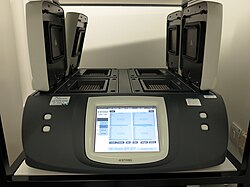
A thermal cycler used for PCR-based molecular testing. (WC)
Utility of molecular pathology
Its utility currently includes:
- Proving clonality, esp. in hematologic malignancies, to help establish a malignant diagnosis.
- Finding recurrent genetic changes - which may be diagnostic, prognostic and suggest a specific therapy.
- Monitor minimal residual disease.
Overview
Molecular pathology can be divided as follows:
| Molecular pathology | |||||||||||||||||||
| Electrophoresis based techniques | Cytogenetics | ||||||||||||||||||
Tabular comparisons
Overview
A simplified overview of molecular pathology:
| Name of technique | Advantages | Disadvantages |
|---|---|---|
| in situ hybridization (ISH) | intermediate resolution - better resolution than karyotyping for the specific target of the given ISH; good way to find gene losses and duplications (one colour) and gene splits and fusions (two colours); can be done on formalin fixed paraffin embedded tissue | target specific (if the target is wrong no information is gained or one is mislead by the negative result); NOT good for "going on a fishing expedition", i.e. looking for changes when one doesn't quite know what is wrong |
| karyotyping | finds large scale changes (gains, losses, rearrangements); good for "going on a fishing expedition", i.e. looking for changes when one doesn't quite know what is wrong | low resolution (completely misses small scale changes); requires fresh tissue/cell culture (as it is based on metaphase nuclei) |
| PCR + sequencing or enzyme digestion and electrophoresis | high resolution (can find very small changes, e.g. base pair substitutions) - considered gold standard; can be done on formalin fixed paraffin embedded tissue | expensive; thus, limited to small regions (target specific); enzyme digestion and electrophoresis is a compromise of sorts where one needs to know something about the expected abnormality; (gene) duplications may be difficult to prove; regions with many repeats may be difficult to sequence |
PCR-based/electrophoresis based techniques
A comparison of common molecular techniques:
| Name of technique | Key elements | Type of change detected | Cost | Other |
|---|---|---|---|---|
| DNA sequencing | PCR, sequencing machine | any (small) DNA change in the genome; does not account for post-transcriptional changes (one cannot definitively infer protein level change) | $$$ | gold standard; will not detect large scale changes unless the break points/fusion regions are sequenced |
| RNA sequencing | reverse transcription PCR, sequencing maching | any change in the mRNA (post-splicing); useful for infering protein level changes | $$$ | slightly less costly than DNA sequencing - as the extrons are not sequenced |
| Restriction fragment length polymorphism (RFLP) | PCR, restriction endonuclease digestion, gel electrophoresis | useful for finding common base pair changes | $$ | value of result depends on RFLP data specific to gene, i.e. knowledge about mutations commonly seen in the gene |
| Amplification-refractory mutation system (ARMS) | PCR with mutation-specific primer, gel electrophoresis | useful for finding a specific known change | $$ | primers can be thought of as a hybridization probe; no mutation-specific hybridization (of primer) --> no PCR product |
| Southern blot | gel electrophoresis, hybridization probe with label | useful for finding a specific known change, quantifying gene copy number | $$$$$ | -rarely done -does not use PCR -considered the gold standard for clonality[1] -most labs consider fresh or frozen tissue a requirement[2] |
Cytogenetics
A comparison of ISH and karyotyping:
| Name of technique | Key elements | Type of change detected | Cost | Other |
|---|---|---|---|---|
| Interphase ISH break apart probe (two colours) | probes label two parts of a (normal) gene; the two markers straddle (common) break points | gene fragmentation consistent with translocation; one may find: gene duplication (or chromosomal duplication), gene loss (or chromosome loss) | $$$$ | can detect translocations - without knowing the specific fusion product |
| Interphase ISH fusion probe (two colours) | probes label different genes (that are not adjacent) | translocation involving the two genes labeled; one may find: gene duplication (or chromosomal duplication), gene loss (or chromosome loss) | $$$$ | can detect one specific translocation |
| Interphase ISH probe (one colour) | probe labels one region (gene) | gene duplication (or chromosomal duplication), gene loss (or chromosome loss) | $$$ | |
| Karyotyping | metaphase nuclei | large scale changes (fusions, deletions, translocations) | $$$$ | gives the "big picture" view of all the (nuclear) DNA |
| Metaphase ISH probe (one colour / two colours) | probe labels one region (one colour) or probes label two parts of a (normal) gene (two colours) or probes label different genes (two colours) | gene duplication, gene loss, translocations | $$$$$ | rarely done; follows karyotyping to better characterize unusual cases; can be thought of as a karyotype and a simultaneous ISH |
PCR-based techniques
General
What?
- Very small changes - submicroscopic.
- Changes in sequence - may be as small as one base pair.
- Used to confirmation chromosomal translocations that are, in clinical practice, usually found with other techniques.
Techniques
- DNA sequencing.
- Real time-PCR, AKA real time-quantitative PCR (RQ-PCR).
- RNA sequencing.
- May be examined after reverse transcription (RNA -> DNA), i.e. RT-PCR.
- Amplification-refractory mutation system (ARMS):[3]
- Technique for finding a (specific) single base change.
- The (PCR) primers are designed bind to the mutated sequence.
- If the mutation is present a PCR product is seen.
- If the mutation is absent no PCR product is seen.
- The (PCR) primers are designed bind to the mutated sequence.
- Technique for finding a (specific) single base change.
- Restriction fragment length polymorphism (RFLP).[4]
- Technique useful for finding a single base change.
- Restriction endonuclease(s), generally, will generate different fragment lengths if nucleotide change is present.
- This techique is most useful if one is looking for a specific (small) genetic change (e.g. F5 Arg534Gln).
- Technique useful for finding a single base change.
Specific tests
A list of tests are found in the Molecular pathology tests article.
DNA & RNA extraction
Other molecular tests
Techniques
- Southern blot.
- DNA quantification.
Key elements:
- Gel electrophoresis.
- Labeling with hybridization probe.
Cytogenetics
Main article: Cytogenetics
This deals with karyotyping and ISH.
Miscellaneous stuff
World protein databank
I can't help think it is ironic that the protein databank goal is to maintain a free and publicly available archive,[7] yet the announcement is in pay-for-access journal (Nature Structual Biology).[8]
Wnt/beta-catenin pathway
Important in hepatoblastomas.[9]
See also
References
- ↑ Medeiros, LJ.; Carr, J. (Dec 1999). "Overview of the role of molecular methods in the diagnosis of malignant lymphomas.". Arch Pathol Lab Med 123 (12): 1189-207. doi:10.1043/0003-9985(1999)1231189:OOTROM2.0.CO;2. PMID 10583924.
- ↑ Reinartz, JJ.; McCormick, SR.; Ikier, DM.; Mellgen, AM.; Bonham, SC.; Strickler, JG.; Mendiola, JR. (Sep 2000). "Immunoglobulin heavy-chain gene rearrangement studies by Southern blot using DNA extracted from formalin-fixed, paraffin-embedded tissue.". Mol Diagn 5 (3): 227-33. doi:10.1054/modi.2000.19808. PMID 11070157.
- ↑ Little S (May 2001). "Amplification-refractory mutation system (ARMS) analysis of point mutations". Curr Protoc Hum Genet Chapter 9: Unit 9.8. doi:10.1002/0471142905.hg0908s07. PMID 18428319.
- ↑ URL: http://www.ncbi.nlm.nih.gov/projects/genome/probe/doc/TechRFLP.shtml. Accessed on: 10 May 2011.
- ↑ Chomczynski P, Sacchi N (2006). "The single-step method of RNA isolation by acid guanidinium thiocyanate-phenol-chloroform extraction: twenty-something years on". Nat Protoc 1 (2): 581–5. doi:10.1038/nprot.2006.83. PMID 17406285.
- ↑ Pikor LA, Enfield KS, Cameron H, Lam WL (2011). "DNA extraction from paraffin embedded material for genetic and epigenetic analyses". J Vis Exp (49). doi:10.3791/2763. PMID 21490570.
- ↑ Worldwide Protein Data Bank. URL: http://www.wwpdb.org/faq.html Accessed on: April 22, 2009.
- ↑ Berman H, Henrick K, Nakamura H (December 2003). "Announcing the worldwide Protein Data Bank". Nat. Struct. Biol. 10 (12): 980. doi:10.1038/nsb1203-980. PMID 14634627.
- ↑ Cotran, Ramzi S.; Kumar, Vinay; Fausto, Nelson; Nelso Fausto; Robbins, Stanley L.; Abbas, Abul K. (2005). Robbins and Cotran pathologic basis of disease (7th ed.). St. Louis, Mo: Elsevier Saunders. pp. 923. ISBN 0-7216-0187-1.