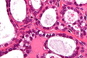
Clear cell carcinoma of the ovary
Jump to navigation
Jump to search
| Clear cell carcinoma of the ovary | |
|---|---|
| Diagnosis in short | |
 Ovarian clear cell carcinoma. H&E stain. | |
|
| |
| Synonyms | ovarian clear cell carcinoma, ovarian clear cell adenocarcinoma, clear cell adenocarcinoma of the ovary |
|
| |
| LM | cystic/tubular architecture (may be papillary and/or solid), clear cytoplasm (may be sparse), hobnail morphology - apical surface larger than basal surface ("nuclei bulge into the lumen"), +/-hyaline droplets, prominent nucleoli |
| LM DDx | serous carcinoma of the ovary, other serous carcinomas, other clear cell tumours (e.g. clear cell renal cell carcinoma), Arias-Stella reaction within endometriosis |
| IHC | HNF-1beta +ve, WT-1 -ve, CK7 +ve, CD10 -ve, CK20 -ve |
| Gross | mass, yellow colour, usu. unilateral |
| Site | ovary - see ovarian tumours |
|
| |
| Associated Dx | endometriosis, pulmonary embolism |
| Prevalence | uncommon |
| Prognosis | poor |
| Clin. DDx | other ovarian tumours, other pelvic and abdominal tumours |
Clear cell carcinoma of the ovary, also ovarian clear cell carcinoma (abbreviated OCCC), is an aggressive malignant tumour of the ovary.
It often forms glands; thus, it is also known as ovarian clear cell adenocarcinoma, and clear cell adenocarcinoma of the ovary.
General
Prognosis:
- Worse prognosis versus other surface epithelial tumours.[1]
Epidemiology:
- Asians appear to be at an increased risk.[2]
- Thought to be related to endometrioid carcinoma.[4]
- Association with endometriosis:[2]
- Increased risk of ovarian clear cell carcinoma ~ 3x.
- Seen in 50-70% of ovarian CCC.
Gross
Features:[2]
- Usually unilateral (unlike serous carcinoma).
- Yellow colour.
Microscopic
Features:
- Cystic/tubular architecture - important low power feature.
- May be papillary and/or solid.
- Clear cells - cytoplasm is clear - key feature. †
- Hobnail morphology - apical surface larger than basal surface.
- "Nuclei bulge into the lumen".
- Hyaline droplets -- common, as in clear cell renal cell carcinoma.
- Eosinophilic bodies within lumen.
- Seen in approximately 1/4 of cases.[2]
- Nucleoli - prominent.
Note:
- † Clear cell adenocarcinoma does not have to have clear cells. Yes, this is stupid; it is like papillary thyroid carcinoma -- which often isn't papillary -- see no truth in names.
- The hobnail morphology is important if this is the case.
DDx:
- Serous carcinoma of the ovary - have larger papillae +/- branching +/-multilayered epithelium, more mitotic activity,[2] more group pleomorphism.
- Serous borderline tumour of the ovary.
- High-grade endometrioid carcinoma of the ovary.
- Metatstatic clear cell carcinoma, esp. clear cell renal cell carcinoma.
- Borderline clear cell lesion of the ovary - very rare; an under sampled carcinoma considered more probable.[2]
- Arias-Stella reaction within endometriosis.
Images
www:
IHC
Panel for high grade serous vs. clear cell:[7]
- ER (+ve in serous),[8] HNF-1beta (+ve in clear cell), WT-1 (+ve in serous).
Others:[2]
- CK7 +ve -- may be focal.
- CK20 -ve.
- CD10 -ve.
- Often +ve in clear cell renal cell carcinoma.
See also
References
- ↑ Hauptmann S, Köbel M (2005). "[Prognostic factors in ovarian carcinoma]" (in German). Verh Dtsch Ges Pathol 89: 92-100. PMID 18035678.
- ↑ 2.0 2.1 2.2 2.3 2.4 2.5 2.6 2.7 Offman, SL.; Longacre, TA. (Sep 2012). "Clear cell carcinoma of the female genital tract (not everything is as clear as it seems).". Adv Anat Pathol 19 (5): 296-312. doi:10.1097/PAP.0b013e31826663b1. PMID 22885379.
- ↑ Duska, LR.; Garrett, L.; Henretta, M.; Ferriss, JS.; Lee, L.; Horowitz, N. (Mar 2010). "When 'never-events' occur despite adherence to clinical guidelines: the case of venous thromboembolism in clear cell cancer of the ovary compared with other epithelial histologic subtypes.". Gynecol Oncol 116 (3): 374-7. doi:10.1016/j.ygyno.2009.10.069. PMID 19922988.
- ↑ Cotran, Ramzi S.; Kumar, Vinay; Fausto, Nelson; Nelso Fausto; Robbins, Stanley L.; Abbas, Abul K. (2005). Robbins and Cotran pathologic basis of disease (7th ed.). St. Louis, Mo: Elsevier Saunders. pp. 1098. ISBN 0-7216-0187-1.
- ↑ Tsuchiya, A.; Sakamoto, M.; Yasuda, J.; Chuma, M.; Ohta, T.; Ohki, M.; Yasugi, T.; Taketani, Y. et al. (Dec 2003). "Expression profiling in ovarian clear cell carcinoma: identification of hepatocyte nuclear factor-1 beta as a molecular marker and a possible molecular target for therapy of ovarian clear cell carcinoma.". Am J Pathol 163 (6): 2503-12. PMID 14633622. http://www.ncbi.nlm.nih.gov/pmc/articles/PMC1892387/?tool=pubmed.
- ↑ Online 'Mendelian Inheritance in Man' (OMIM) 189907
- ↑ Köbel M, Kalloger SE, Carrick J, et al. (January 2009). "A limited panel of immunomarkers can reliably distinguish between clear cell and high-grade serous carcinoma of the ovary". Am. J. Surg. Pathol. 33 (1): 14–21. doi:10.1097/PAS.0b013e3181788546. PMID 18830127.
- ↑ DeLair, D.; Oliva, E.; Köbel, M.; Macias, A.; Gilks, CB.; Soslow, RA. (Jan 2011). "Morphologic spectrum of immunohistochemically characterized clear cell carcinoma of the ovary: a study of 155 cases.". Am J Surg Pathol 35 (1): 36-44. doi:10.1097/PAS.0b013e3181ff400e. PMID 21164285.


