Difference between revisions of "Myxopapillary ependymoma"
Jump to navigation
Jump to search
Jensflorian (talk | contribs) m (→Images: touch) |
m (fix stain decript) |
||
| Line 3: | Line 3: | ||
| Image = Myxopapillary_ependymoma_-_very_high_mag.jpg | | Image = Myxopapillary_ependymoma_-_very_high_mag.jpg | ||
| Width = | | Width = | ||
| Caption = Myxopapillary ependymoma. [[ | | Caption = Myxopapillary ependymoma. [[HPS stain]]. | ||
| Synonyms = | | Synonyms = | ||
| Micro = Papillary tumor cells around vascular myxoid matrix | | Micro = Papillary tumor cells around vascular myxoid matrix | ||
Revision as of 12:56, 28 April 2015
| Myxopapillary ependymoma | |
|---|---|
| Diagnosis in short | |
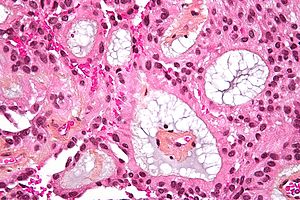
 Myxopapillary ependymoma. HPS stain. | |
|
| |
| LM | Papillary tumor cells around vascular myxoid matrix |
| Subtypes | subtype of ependymoma |
| LM DDx | chordoma, myxoid chondrosarcoma, paraganglioma, papillary adenocarcinoma |
| Stains | Alcian blue +ve |
| IHC | GFAP +ve |
| Site | usually lumbar spinal cord |
|
| |
| Prognosis | good (WHO Grade I) |
Mxyopapillary Ependymoma, is a low-grade Ependymoma. It is nearly always associated with cauda equina and filum terminale.
General
- Low-grade ependymoma - WHO Grade I by definition.
- Classically in the spinal cord of adults.
- Approx 9-13% of all ependymal tumors.[1]
- Associated with back pain.
- Enhancing mass
Gross
- Soft.
- Gray.
- Discrete masses.
- Often encapsulated.
- Subtotal resected tumors may spread throughout the neuraxis.
Microscopic
Features:
- Papillary appearance.
- Perivascular pseudorosettes:
- Cuboidal to elongated tumor cells.
- Radially arranged around vascular cores.
- Myxoid material surround blood vessels.
- Microcysts.
- Low mitotic activity.
Note: Cases with extensive sclerosis may mimic degenerative changes. [2]
DDx:
- Chordoma.
- Myxoid chondrosarcoma.
- Paraganglioma.
- Papillary adenocarcinoma.
Images
- Myxopapillary ependymoma - high mag. (WC).
- Myxopapillary ependymoma (bmj.com) - part of careers.bmj.com article on paediatric pathology.
- Myxopapillary ependymoma - cytology (WC).
- Myxopapillary ependymoma - several images (upmc.edu).
IHC
- GFAP+ve.
- S-100+ve.
- MIB-1 <1%.
Molecular
- Poorly characterized.
- No consistent abberations.
See also
References
- ↑ Schiffer, D.; Chiò, A.; Giordana, MT.; Migheli, A.; Palma, L.; Pollo, B.; Soffietti, R.; Tribolo, A. (Aug 1991). "Histologic prognostic factors in ependymoma.". Childs Nerv Syst 7 (4): 177-82. PMID 1933913.
- ↑ Schittenhelm, J.; Becker, R.; Capper, D.; Meyermann, R.; Iglesias-Rozas, JR.; Kaminsky, J.; Mittelbronn, M.. "The clinico-surgico-pathological spectrum of myxopapillary ependymomas--report of four unusal cases and review of the literature.". Clin Neuropathol 27 (1): 21-8. PMID 18257471.


