Difference between revisions of "Follicular thyroid adenoma"
Jump to navigation
Jump to search
(redirect w/ cat.) |
|||
| (7 intermediate revisions by the same user not shown) | |||
| Line 1: | Line 1: | ||
{{ Infobox diagnosis | |||
| Name = {{PAGENAME}} | |||
| Image = Follicular adenoma -- low mag.jpg | |||
| Width = | |||
| Caption = Follicular adenoma. [[H&E stain]]. | |||
| Synonyms = follicular adenoma | |||
| Micro = cellular appearance (low magnification), microfollicles, thick fibrous capsule without invasion, negative for nuclear features of papillary thyroid carcinoma | |||
| Subtypes = | |||
| LMDDx = [[thyroid gland nodular hyperplasia]], [[follicular thyroid carcinoma]], [[noninvasive follicular thyroid neoplasm with papillary-like nuclear features]] (NIFTP), [[Papillary thyroid carcinoma follicular variant]] | |||
| Stains = | |||
| IHC = | |||
| EM = | |||
| Molecular = | |||
| IF = | |||
| Gross = lesion with thick capsule | |||
| Grossing = | |||
| Staging = | |||
| Site = [[thyroid gland]] | |||
| Assdx = | |||
| Syndromes = | |||
| Clinicalhx = | |||
| Signs = thyroid mass | |||
| Symptoms = | |||
| Prevalence = uncommon | |||
| Bloodwork = | |||
| Rads = | |||
| Endoscopy = | |||
| Prognosis = benign | |||
| Other = | |||
| ClinDDx = [[follicular carcinoma]], other thyroid tumours | |||
| Tx = excision to exclude carcinoma | |||
}} | |||
'''Follicular thyroid adenoma''', abbreviated '''FTA''', is a benign lesion of the [[thyroid gland]]. | |||
==General== | |||
*Most common neoplasm of thyroid.<ref name=Ref_EP51>{{Ref EP|51}}</ref> | |||
*Encapusled lesion (surrounded by fibrous capsule). | |||
*Cannot be diagnosed on [[thyroid cytopathology|thyroid FNA]], as one cannot exclude invasion through the capsule without examining all of it. | |||
==Gross== | |||
*Thick capsule. | |||
Notes: | |||
*The entire capsule should be submitted.<ref>SR. 17 January 2011.</ref> | |||
**A good start for most thyroid specimens with a thick capsule is 10 blocks. | |||
===Images=== | |||
<gallery> | |||
Image: Follicular Adenoma of the Thyroid Gland (5186991355).jpg | FTA. (WC/Euthman) | |||
</gallery> | |||
==Microsopic== | |||
Features: | |||
*Cellular. | |||
*Thick capsule - '''key feature'''. | |||
Negatives. | |||
*No invasion of the capsule - see [[follicular thyroid carcinoma]]. | |||
*No nuclear features suggestive of [[papillary thyroid carcinoma]]. | |||
DDx: | |||
*[[Thyroid gland nodular hyperplasia]] with an encapsulated nodule - not as cellular. | |||
*[[Follicular thyroid carcinoma]]. | |||
*[[Noninvasive follicular thyroid neoplasm with papillary-like nuclear features]] (NIFTP). | |||
*[[Papillary thyroid carcinoma follicular variant]]. | |||
===Images=== | |||
<gallery> | |||
Image: Follicular adenoma -- extremely low mag.jpg | FA - extremely low mag. (WC) | |||
Image: Follicular adenoma -- very low mag.jpg | FA - very low mag. (WC) | |||
Image: Follicular adenoma -- low mag.jpg | FA - low mag. (WC) | |||
Image: Follicular adenoma -- intermed mag.jpg | FA - intermed. mag. (WC) | |||
Image: Follicular adenoma -- high mag.jpg | FA - high mag. (WC) | |||
Image: Follicular adenoma -- very high mag.jpg | FA - very high mag. (WC) | |||
</gallery> | |||
==Sign out== | |||
<pre> | |||
Left Hemithyroid, Hemithyroidectomy: | |||
- Follicular adenoma. | |||
- Parathyroid gland. | |||
- Five benign lymph nodes (0/5). | |||
- NEGATIVE for evidence of malignancy. | |||
</pre> | |||
===Block letters=== | |||
<pre> | |||
LEFT THYROID, SUPERIOR POLE, EXCISION: | |||
- FOLLICULAR ADENOMA, MAXIMAL DIMENSION 5 MM. | |||
- LYMPHOCYTIC THYROIDITIS. | |||
- NODULAR HYPERPLASIA. | |||
- NEGATIVE FOR MALIGNANCY. | |||
</pre> | |||
===Micro=== | |||
The section shows a well-circumscribed lesion encapsulated by a thick fibrous capsule (~0.4 mm thick). | |||
The lesions consists of microfollicles with a dense appearing colloid. The nuclei have round regular nuclear membranes. Small indistinct nucleoli are seen at high power. | |||
Focally, the lesional cells overlap. However, the chromatin is not cleared. Nuclear grooves are not readily apparent and nuclear pseudoinclusions are not readily identified. | |||
==See also== | |||
*[[Thyroid gland]]. | |||
==References== | |||
{{Reflist|1}} | |||
[[Category:Diagnosis]] | [[Category:Diagnosis]] | ||
[[Category:Thyroid gland]] | |||
Latest revision as of 03:41, 10 June 2016
| Follicular thyroid adenoma | |
|---|---|
| Diagnosis in short | |
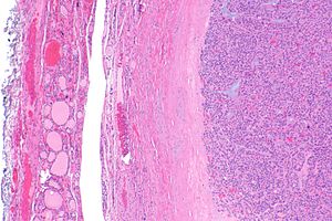
 Follicular adenoma. H&E stain. | |
|
| |
| Synonyms | follicular adenoma |
|
| |
| LM | cellular appearance (low magnification), microfollicles, thick fibrous capsule without invasion, negative for nuclear features of papillary thyroid carcinoma |
| LM DDx | thyroid gland nodular hyperplasia, follicular thyroid carcinoma, noninvasive follicular thyroid neoplasm with papillary-like nuclear features (NIFTP), Papillary thyroid carcinoma follicular variant |
| Gross | lesion with thick capsule |
| Site | thyroid gland |
|
| |
| Signs | thyroid mass |
| Prevalence | uncommon |
| Prognosis | benign |
| Clin. DDx | follicular carcinoma, other thyroid tumours |
| Treatment | excision to exclude carcinoma |
Follicular thyroid adenoma, abbreviated FTA, is a benign lesion of the thyroid gland.
General
- Most common neoplasm of thyroid.[1]
- Encapusled lesion (surrounded by fibrous capsule).
- Cannot be diagnosed on thyroid FNA, as one cannot exclude invasion through the capsule without examining all of it.
Gross
- Thick capsule.
Notes:
- The entire capsule should be submitted.[2]
- A good start for most thyroid specimens with a thick capsule is 10 blocks.
Images
Microsopic
Features:
- Cellular.
- Thick capsule - key feature.
Negatives.
- No invasion of the capsule - see follicular thyroid carcinoma.
- No nuclear features suggestive of papillary thyroid carcinoma.
DDx:
- Thyroid gland nodular hyperplasia with an encapsulated nodule - not as cellular.
- Follicular thyroid carcinoma.
- Noninvasive follicular thyroid neoplasm with papillary-like nuclear features (NIFTP).
- Papillary thyroid carcinoma follicular variant.
Images
Sign out
Left Hemithyroid, Hemithyroidectomy:
- Follicular adenoma.
- Parathyroid gland.
- Five benign lymph nodes (0/5).
- NEGATIVE for evidence of malignancy.
Block letters
LEFT THYROID, SUPERIOR POLE, EXCISION:
- FOLLICULAR ADENOMA, MAXIMAL DIMENSION 5 MM.
- LYMPHOCYTIC THYROIDITIS.
- NODULAR HYPERPLASIA.
- NEGATIVE FOR MALIGNANCY.
Micro
The section shows a well-circumscribed lesion encapsulated by a thick fibrous capsule (~0.4 mm thick).
The lesions consists of microfollicles with a dense appearing colloid. The nuclei have round regular nuclear membranes. Small indistinct nucleoli are seen at high power.
Focally, the lesional cells overlap. However, the chromatin is not cleared. Nuclear grooves are not readily apparent and nuclear pseudoinclusions are not readily identified.
See also
References
- ↑ Thompson, Lester D. R. (2006). Endocrine Pathology: A Volume in Foundations in Diagnostic Pathology Series (1st ed.). Churchill Livingstone. pp. 51. ISBN 978-0443066856.
- ↑ SR. 17 January 2011.


