Difference between revisions of "High-grade astrocytoma with piloid features"
Jump to navigation
Jump to search
Jensflorian (talk | contribs) (Update infobox) |
Jensflorian (talk | contribs) (HGAP) |
||
| Line 31: | Line 31: | ||
}} | }} | ||
'''High-grade astrocytoma with piloid features''' is a rare [[Glioma|glial]] tumor which often requires methylation analyis to secure diagnosis. There is currently no definitive grading, but clinical behaviour suggests WHO CNS grade 3. | '''High-grade astrocytoma with piloid features''' (HGAP) is a rare [[Glioma|glial]] tumor which often requires methylation analyis to secure diagnosis. There is currently no definitive grading, but clinical behaviour suggests WHO CNS grade 3. | ||
| Line 54: | Line 54: | ||
==Images== | ==Images== | ||
<gallery> | <gallery> | ||
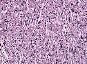
Image:Pilocytic astrocytoma with anaplastic features.jpg | | Image:Pilocytic astrocytoma with anaplastic features.jpg | HGAP, [[FFPE]] specimen, HE (WC). | ||
Image:Pilocytic astrocytoma with anaplastic features mimicking focally a GBM.jpg | Vacular proliferations and necrosis in | Image:Pilocytic astrocytoma with anaplastic features mimicking focally a GBM.jpg | Vacular proliferations and necrosis in HGAP mimicking [[GBM]] (WC). | ||
</gallery> | </gallery> | ||
Latest revision as of 13:54, 17 October 2022
| High-grade astrocytoma with piloid features | |
|---|---|
| Diagnosis in short | |
 High-grade astrocytoma with piloid features. H&E stain. | |
| LM DDx | astrocytoma, PXA, glioblastoma |
| Stains | PAS-D +ve (eosinophilic granular bodies) |
| IHC | GFAP +ve |
| Gross | usually cerebellar |
| Site | brain - usu. cerebellum |
|
| |
| Prevalence | very rare - esp. in children |
| Prognosis | poor (analog to WHO Grade III) |
High-grade astrocytoma with piloid features (HGAP) is a rare glial tumor which often requires methylation analyis to secure diagnosis. There is currently no definitive grading, but clinical behaviour suggests WHO CNS grade 3.
Clinical
- Rare (1-3% of all brain tumor in adults).
- Usu. posterior fossa (75%).
- Supratentorial or spinal locations possible.
- Imaging may be similiar to Glioblastoma.
- 5-year OS: 50%.
- De novo cases in the setting of neurofibromatosis 1 reported.
- Tumors may have history of previous radiation.
Histology
- Often so variable, so molecular testing is essential to secure diagnosis.
- Astrocytic nature of tumor cells.
- Frequent mitoses.
- Elongated glial tumor cell processes ("piloid").
- Rosenthal fibers or eosinophilic granular bodies.
- Perivascular lymphocytic cuffing.
- Necrosis may be present.
Images
IHC
- GFAP+ve.
- ATRX: nuclear loss in approx. 40%.
Molecular
- DNA Methylation profile of high-grade astrocytoma with piloid features is essential for diagnosis.
- MAPK genes often altered (NF1, BRAF fusion, FGFR1, KRAS).
- CDKN2A/B homozygous deletion.
- CDK4 amplification.
- TERT promotor mutations are rare.
- Absence of IDH1/2 hotspot mutation.
Prognosis
- Poor, but better than conventional glioblastoma.[1]
- Prognostic unfavourable paramters include: Necrosis, Mitoses and previous radiation.
DDx:
See also
- ↑ Rodriguez, FJ.; Scheithauer, BW.; Burger, PC.; Jenkins, S.; Giannini, C. (Feb 2010). "Anaplasia in pilocytic astrocytoma predicts aggressive behavior.". Am J Surg Pathol 34 (2): 147-60. doi:10.1097/PAS.0b013e3181c75238. PMID 20061938.

