Difference between revisions of "Sarcina"
Jump to navigation
Jump to search

(→Microscopic: +SO) |
(+uring) |
||
| Line 41: | Line 41: | ||
Image: Gastritis with Sarcina - a1 - very high mag.jpg | Sarcina - very high mag. | Image: Gastritis with Sarcina - a1 - very high mag.jpg | Sarcina - very high mag. | ||
</gallery> | </gallery> | ||
====Case 2==== | |||
<gallery> | |||
Image: Sarcina - urine -- intermed mag.jpg | Sarcina - urine cytology - intermed. mag. | |||
Image: Sarcina - urine -- high mag.jpg | Sarcina - urine cytology - high mag. | |||
Image: Sarcina - urine - alt -- high mag.jpg | Sarcina - urine cytology - high mag. | |||
Image: Sarcina - urine -- very high mag.jpg | Sarcina - urine cytology - very high mag. | |||
Image: Sarcina - urine -- extremely high mag.jpg | Sarcina - urine cytology - extremely high mag. | |||
</gallery> | |||
====www==== | ====www==== | ||
*[http://www.ncbi.nlm.nih.gov/pmc/articles/PMC3193598/figure/F2/ Sarcina (nih.gov)]. | *[http://www.ncbi.nlm.nih.gov/pmc/articles/PMC3193598/figure/F2/ Sarcina (nih.gov)]. | ||
Latest revision as of 23:36, 3 November 2024

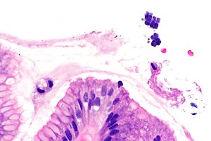
Micrograph showing Sarcina. H&E stain. (WC/Librepath)
Sarcina organisms are occasionally seen in the gastrointestinal tract. They are reported in urine.[1]
General
- Probably a commensal organism.[2]
- Gram positive coccus.[1]
- Uncommonly seen.
- Associated with gastroparesis,[2] as may be seen in diabetes mellitus.
Described complications:[2]
- Gastric perforation.
- Emphysematous gastritis.
Notes:
- Sarcina ventriculi = Gram-positive organism.[2]
Gross
Features:
- Erythema.
- +/-Gastric ulcer.
- +/-Retained food.
Microscopic
Features:[3]
- Small essentially spherical micro-organisms.
- Each micro-organism 1.8-3 micrometres - purple on H&E stain.
- Arranged in clusters of 4, 8 or more - classically in a tetrad - key feature.
DDx:
- Micrococcus - form larger clusters.[3]
Images
Case 1
Case 2
www
Sign out
Urine cytology
Abundant micro-organisms compatible with Sarcina, see comment.
Acute inflammation.
NEGATIVE for High-Grade Urothelial Carcinoma.

Comment:
The micro-organisms are arranged in distinctive tetrads, as is typical for Sarcina.
This is an uncommon finding.[1] Treatment should be considered within the clinical context.

1. Bommannan K, Gaspar BL, Sachdeva MUS. BMJ Case Rep. 2016 Oct 13;2016:bcr2016216991. doi:10.1136/bcr-2016-216991
https://www.ncbi.nlm.nih.gov/pmc/articles/PMC5073697/pdf/bcr-2016-216991.pdf
See also
References
- ↑ 1.0 1.1 Bommannan K, Gaspar BL, Sachdeva MU (October 2016). "Pathogenic Sarcina in urine". BMJ Case Rep 2016. doi:10.1136/bcr-2016-216991. PMC 5073697. PMID 27737866. https://www.ncbi.nlm.nih.gov/pmc/articles/PMC5073697/.
- ↑ 2.0 2.1 2.2 2.3 2.4 Ratuapli, SK.; Lam-Himlin, DM.; Heigh, RI. (2013). "Sarcina ventriculi of the stomach: a case report.". World J Gastroenterol 19 (14): 2282-5. doi:10.3748/wjg.v19.i14.2282. PMID 23599657.
- ↑ 3.0 3.1 Lam-Himlin, D.; Tsiatis, AC.; Montgomery, E.; Pai, RK.; Brown, JA.; Razavi, M.; Lamps, L.; Eshleman, JR. et al. (Nov 2011). "Sarcina organisms in the gastrointestinal tract: a clinicopathologic and molecular study.". Am J Surg Pathol 35 (11): 1700-5. doi:10.1097/PAS.0b013e31822911e6. PMID 21997690.


