Difference between revisions of "Metanephric adenoma"
Jump to navigation
Jump to search
| (3 intermediate revisions by the same user not shown) | |||
| Line 1: | Line 1: | ||
{{ Infobox diagnosis | {{ Infobox diagnosis | ||
| Name = {{PAGENAME}} | | Name = {{PAGENAME}} | ||
| Image = Metanephric_adenoma_-_very_high_mag.jpg | | Image = Metanephric_adenoma_-_very_high_mag.jpg | ||
| Width = | | Width = | ||
| Caption = Metanephric adenoma. [[H&E stain]]. | | Caption = Metanephric adenoma. [[H&E stain]]. | ||
| Line 7: | Line 7: | ||
| Micro = small uniform cells with fine chromatin, no apparent [[nucleolus]], and a relatively smooth nuclear membrane; variable architecture - may be sheets, ductal or micropapillary, +/-[[psammoma bodies]] | | Micro = small uniform cells with fine chromatin, no apparent [[nucleolus]], and a relatively smooth nuclear membrane; variable architecture - may be sheets, ductal or micropapillary, +/-[[psammoma bodies]] | ||
| Subtypes = | | Subtypes = | ||
| LMDDx = [[Wilms tumour]], [[papillary renal cell carcinoma]] (type 1) | | LMDDx = [[Wilms tumour]], [[papillary renal cell carcinoma]] (type 1), [[renal mucinous tubular and spindle cell carcinoma]] | ||
| Stains = | | Stains = | ||
| IHC = WT-1 +ve, CD57 +ve, AMACR -ve, CD56 -ve | | IHC = WT-1 +ve, CD57 +ve, AMACR -ve, CD56 -ve | ||
| Line 43: | Line 43: | ||
==Gross== | ==Gross== | ||
*Sharply circumscribed. | *Sharply circumscribed. | ||
*Not encapsulated. | *Not encapsulated.<ref name=pmid23551615>{{Cite journal | last1 = Mantoan Padilha | first1 = M. | last2 = Billis | first2 = A. | last3 = Allende | first3 = D. | last4 = Zhou | first4 = M. | last5 = Magi-Galluzzi | first5 = C. | title = Metanephric adenoma and solid variant of papillary renal cell carcinoma: common and distinctive features. | journal = Histopathology | volume = 62 | issue = 6 | pages = 941-53 | month = May | year = 2013 | doi = 10.1111/his.12106 | PMID = 23551615 }}</ref> | ||
==Microscopic== | ==Microscopic== | ||
| Line 60: | Line 60: | ||
**Mitoses (rare in ''metanephric adenoma''). | **Mitoses (rare in ''metanephric adenoma''). | ||
*[[Papillary renal cell carcinoma|Papillary RCC]].<ref name=Ref_WMSP284>{{Ref WMSP|284}}</ref> | *[[Papillary renal cell carcinoma|Papillary RCC]].<ref name=Ref_WMSP284>{{Ref WMSP|284}}</ref> | ||
*[[Renal mucinous tubular and spindle cell carcinoma]]. | |||
*Other [[small round cell tumours]], e.g. [[small cell carcinoma]]. | *Other [[small round cell tumours]], e.g. [[small cell carcinoma]]. | ||
Latest revision as of 17:06, 17 December 2017
| Metanephric adenoma | |
|---|---|
| Diagnosis in short | |
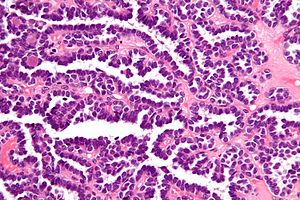
 Metanephric adenoma. H&E stain. | |
|
| |
| LM | small uniform cells with fine chromatin, no apparent nucleolus, and a relatively smooth nuclear membrane; variable architecture - may be sheets, ductal or micropapillary, +/-psammoma bodies |
| LM DDx | Wilms tumour, papillary renal cell carcinoma (type 1), renal mucinous tubular and spindle cell carcinoma |
| IHC | WT-1 +ve, CD57 +ve, AMACR -ve, CD56 -ve |
| Site | kidney - see kidney tumours |
|
| |
| Clinical history | usu. adults, occasionally children |
| Prevalence | uncommon |
| Prognosis | good, benign |
Metanephric adenoma is a benign tumour of the kidney.
It should not be confused mesonephric adenoma, another term for nephrogenic adenoma.
This can be remembered as follows: metanephric adenoma is a tumour.
General
- Benign.
- Afflicts adults and occasionally children.
- May be associated with polycythemia.[1]
Gross
- Sharply circumscribed.
- Not encapsulated.[2]
Microscopic
Features:[3]
- Small uniform cells with:
- Fine chromatin.
- No apparent nucleolus.
- A relatively smooth nuclear membrane.
- Variable architecture - may be sheets, ductal or micropapillary.
- +/-Psammoma bodies.
DDx:
- Epithelioid nephroblastoma (Wilms tumour) - these typically have:
- Irregular nuclear membrane.
- Nucleoli.
- Mitoses (rare in metanephric adenoma).
- Papillary RCC.[3]
- Renal mucinous tubular and spindle cell carcinoma.
- Other small round cell tumours, e.g. small cell carcinoma.
Images
www:
IHC
- WT-1 +ve.[4]
- CD57 +ve.[4]
- AMACR -ve.[4]
- CD56 -ve.[5]
- Positive in Wilms tumour[5] and small cell carcinoma.
- CK7 +ve (focal).[5]
- TTF-1 -ve.[citation needed]
Sign out
Left Kidney, Total Nephrectomy:
- Metanephric adenoma, see comment.

COMMENT:
The tumour stains as follows:
POSITIVE: WT1, CD57, PAX8.
NEGATIVE: CD56, TTF-1, AMACR.
See also
References
- ↑ Le Nué, R.; Marcellin, L.; Ripepi, M.; Henry, C.; Kretz, JM.; Geiss, S. (Aug 2011). "Conservative treatment of metanephric adenoma. A case report and review of the literature.". J Pediatr Urol 7 (4): 399-403. doi:10.1016/j.jpurol.2010.09.010. PMID 21220212.
- ↑ Mantoan Padilha, M.; Billis, A.; Allende, D.; Zhou, M.; Magi-Galluzzi, C. (May 2013). "Metanephric adenoma and solid variant of papillary renal cell carcinoma: common and distinctive features.". Histopathology 62 (6): 941-53. doi:10.1111/his.12106. PMID 23551615.
- ↑ 3.0 3.1 Humphrey, Peter A; Dehner, Louis P; Pfeifer, John D (2008). The Washington Manual of Surgical Pathology (1st ed.). Lippincott Williams & Wilkins. pp. 284. ISBN 978-0781765275.
- ↑ 4.0 4.1 4.2 Watanabe, S.; Naganuma, H.; Shimizu, M.; Ota, S.; Murata, S.; Nihei, N.; Matsushima, J.; Mikami, S. et al. (2013). "Adult nephroblastoma with predominant epithelial component: a differential diagnostic candidate of papillary renal cell carcinoma and metanephric adenoma-report of three cases.". Case Rep Pathol 2013: 675875. doi:10.1155/2013/675875. PMID 24083046.
- ↑ 5.0 5.1 5.2 Muir, TE.; Cheville, JC.; Lager, DJ. (Oct 2001). "Metanephric adenoma, nephrogenic rests, and Wilms' tumor: a histologic and immunophenotypic comparison.". Am J Surg Pathol 25 (10): 1290-6. PMID 11688464.
.


