Difference between revisions of "Hyaline globules"
Jump to navigation
Jump to search
m (→Tumours: w) |
|||
| (11 intermediate revisions by 2 users not shown) | |||
| Line 1: | Line 1: | ||
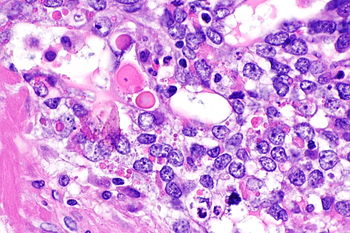
[[Image:Yolk sac tumour with hyaline bodies -- very high mag.jpg|thumb|350px|right|Hyaline globules in a [[yolk sac tumour]]. [[H&E stain]].]] | |||
'''Hyaline globules''', also '''hyaline bodies''', are a common non-specific histomorphologic feature that can be useful in formulating a differential diagnosis. | '''Hyaline globules''', also '''hyaline bodies''', are a common non-specific histomorphologic feature that can be useful in formulating a differential diagnosis. | ||
| Line 11: | Line 12: | ||
==Tumours== | ==Tumours== | ||
Gynecologic: | |||
*[[Clear cell carcinoma]] (CCC). | *[[Clear cell carcinoma]] (CCC). | ||
*[[ | *[[Yolk sac tumour]], hepatoid pattern.<ref>URL: [http://webpathology.com/image.asp?case=34&n=6 http://webpathology.com/image.asp?case=34&n=6]. Accessed on: March 8, 2010.</ref> | ||
Gastrointestinal: | |||
*[[Solid pseudopapillary tumour]].<ref name=pmid18708424>{{cite journal |author=Serra S, Chetty R |title=Revision 2: an immunohistochemical approach and evaluation of solid pseudopapillary tumour of the pancreas |journal=J. Clin. Pathol. |volume=61 |issue=11 |pages=1153–9 |year=2008 |month=November |pmid=18708424 |doi=10.1136/jcp.2008.057828 |url=http://jcp.bmj.com/content/61/11/1153}}</ref> | *[[Solid pseudopapillary tumour]].<ref name=pmid18708424>{{cite journal |author=Serra S, Chetty R |title=Revision 2: an immunohistochemical approach and evaluation of solid pseudopapillary tumour of the pancreas |journal=J. Clin. Pathol. |volume=61 |issue=11 |pages=1153–9 |year=2008 |month=November |pmid=18708424 |doi=10.1136/jcp.2008.057828 |url=http://jcp.bmj.com/content/61/11/1153}}</ref> | ||
*Pancreatic endocrine tumour.<ref name=pmid18708424>{{cite journal |author=Serra S, Chetty R |title=Revision 2: an immunohistochemical approach and evaluation of solid pseudopapillary tumour of the pancreas |journal=J. Clin. Pathol. |volume=61 |issue=11 |pages=1153–9 |year=2008 |month=November |pmid=18708424 |doi=10.1136/jcp.2008.057828 |url=http://jcp.bmj.com/content/61/11/1153}}</ref> | *Pancreatic endocrine tumour.<ref name=pmid18708424>{{cite journal |author=Serra S, Chetty R |title=Revision 2: an immunohistochemical approach and evaluation of solid pseudopapillary tumour of the pancreas |journal=J. Clin. Pathol. |volume=61 |issue=11 |pages=1153–9 |year=2008 |month=November |pmid=18708424 |doi=10.1136/jcp.2008.057828 |url=http://jcp.bmj.com/content/61/11/1153}}</ref> | ||
*[[Hepatocellular carcinoma]] - very common.<ref name=pmid11026104>{{Cite journal | last1 = Nayar | first1 = R. | last2 = Bourtsos | first2 = E. | last3 = DeFrias | first3 = DV. | title = Hyaline globules in renal cell carcinoma and hepatocellular carcinoma. A clue or a diagnostic pitfall on fine-needle aspiration? | journal = Am J Clin Pathol | volume = 114 | issue = 4 | pages = 576-82 | month = Oct | year = 2000 | doi = 10.1309/F4TU-6AFE-R7NU-39Y3 | PMID = 11026104 }}</ref> | *[[Hepatocellular carcinoma]] - very common.<ref name=pmid11026104>{{Cite journal | last1 = Nayar | first1 = R. | last2 = Bourtsos | first2 = E. | last3 = DeFrias | first3 = DV. | title = Hyaline globules in renal cell carcinoma and hepatocellular carcinoma. A clue or a diagnostic pitfall on fine-needle aspiration? | journal = Am J Clin Pathol | volume = 114 | issue = 4 | pages = 576-82 | month = Oct | year = 2000 | doi = 10.1309/F4TU-6AFE-R7NU-39Y3 | PMID = 11026104 }}</ref> | ||
Other: | |||
*[[Renal cell carcinoma]].<ref name=pmid11026104/> | *[[Renal cell carcinoma]].<ref name=pmid11026104/> | ||
*[[Kaposi sarcoma]] (KS). | |||
* Secretory [[Meningioma]].<ref>{{Cite journal | last1 = Budka | first1 = H. | title = Hyaline inclusions (Pseudopsammoma bodies) in meningiomas: immunocytochemical demonstration of epithel-like secretion of secretory component and immunoglobulins A and M. | journal = Acta Neuropathol | volume = 56 | issue = 4 | pages = 294-8 | month = | year = 1982 | doi = | PMID = 6283779 }}</ref> | |||
*[[Lung adenocarcinoma]].<ref>{{cite journal |authors=Haninger DM, Kloecker GH, Bousamra Ii M, Nowacki MR, Slone SP |title=Hepatoid adenocarcinoma of the lung: report of five cases and review of the literature |journal=Mod Pathol |volume=27 |issue=4 |pages=535–42 |date=April 2014 |pmid=24030743 |doi=10.1038/modpathol.2013.170 |url=}}</ref> | |||
**Estimated to be present in 6% of lung adenocarcinomas (in cohort of 100 adenocarcinomas).<ref>{{cite journal |authors=Scroggs MW, Roggli VL, Fraire AE, Sanfilippo F |title=Eosinophilic intracytoplasmic globules in pulmonary adenocarcinomas: a histochemical, immunohistochemical, and ultrastructural study of six cases |journal=Hum Pathol |volume=20 |issue=9 |pages=845–9 |date=September 1989 |pmid=2476374 |doi=10.1016/0046-8177(89)90095-6 |url=}}</ref> | |||
===Images=== | |||
<gallery> | |||
Image:Ovarian_clear_cell_carcinoma_-a-_very_high_mag.jpg | Hyaline globules in CCC - very high mag. (WC) | |||
Image:Kaposi_sarcoma_high_mag.jpg | Hyaline globules in KS - high mag. (WC) | |||
Image:Solid_pseudopapillary_tumour_-_very_high_mag.jpg | Hyaline globules in solid pseudopapillary tumour - very high mag. (WC) | |||
File:Secretory meningioma HE x200.jpg | Hyaline globules (pseudopsammoma bodies) in secretory meningioma (WC/jensflorian) | |||
</gallery> | |||
==See also== | |||
*[ | *[[Psammoma bodies]]. | ||
==References== | ==References== | ||
Latest revision as of 15:34, 7 May 2022
Hyaline globules, also hyaline bodies, are a common non-specific histomorphologic feature that can be useful in formulating a differential diagnosis.
They can be seen in benign and malignant tissue.
Microscopic
Features:
- Eosinophilic (pink) round bodies ~ typically 4-10 micrometers in diameter.
Benign
- Ectopic decidua.[1]
Tumours
Gynecologic:
- Clear cell carcinoma (CCC).
- Yolk sac tumour, hepatoid pattern.[2]
Gastrointestinal:
- Solid pseudopapillary tumour.[3]
- Pancreatic endocrine tumour.[3]
- Hepatocellular carcinoma - very common.[4]
Other:
- Renal cell carcinoma.[4]
- Kaposi sarcoma (KS).
- Secretory Meningioma.[5]
- Lung adenocarcinoma.[6]
- Estimated to be present in 6% of lung adenocarcinomas (in cohort of 100 adenocarcinomas).[7]
Images
See also
References
- ↑ Dharan M (September 2009). "Hyaline globules in ectopic decidua in a pregnant woman with cervical squamous cell carcinoma". Diagn. Cytopathol. 37 (9): 696–8. doi:10.1002/dc.21113. PMID 19526574.
- ↑ URL: http://webpathology.com/image.asp?case=34&n=6. Accessed on: March 8, 2010.
- ↑ 3.0 3.1 Serra S, Chetty R (November 2008). "Revision 2: an immunohistochemical approach and evaluation of solid pseudopapillary tumour of the pancreas". J. Clin. Pathol. 61 (11): 1153–9. doi:10.1136/jcp.2008.057828. PMID 18708424. http://jcp.bmj.com/content/61/11/1153.
- ↑ 4.0 4.1 Nayar, R.; Bourtsos, E.; DeFrias, DV. (Oct 2000). "Hyaline globules in renal cell carcinoma and hepatocellular carcinoma. A clue or a diagnostic pitfall on fine-needle aspiration?". Am J Clin Pathol 114 (4): 576-82. doi:10.1309/F4TU-6AFE-R7NU-39Y3. PMID 11026104.
- ↑ Budka, H. (1982). "Hyaline inclusions (Pseudopsammoma bodies) in meningiomas: immunocytochemical demonstration of epithel-like secretion of secretory component and immunoglobulins A and M.". Acta Neuropathol 56 (4): 294-8. PMID 6283779.
- ↑ Haninger DM, Kloecker GH, Bousamra Ii M, Nowacki MR, Slone SP (April 2014). "Hepatoid adenocarcinoma of the lung: report of five cases and review of the literature". Mod Pathol 27 (4): 535–42. doi:10.1038/modpathol.2013.170. PMID 24030743.
- ↑ Scroggs MW, Roggli VL, Fraire AE, Sanfilippo F (September 1989). "Eosinophilic intracytoplasmic globules in pulmonary adenocarcinomas: a histochemical, immunohistochemical, and ultrastructural study of six cases". Hum Pathol 20 (9): 845–9. doi:10.1016/0046-8177(89)90095-6. PMID 2476374.


