Difference between revisions of "Granulomatosis with polyangiitis"
Jump to navigation
Jump to search
(split out) |
|||
| (9 intermediate revisions by the same user not shown) | |||
| Line 1: | Line 1: | ||
'''Granulomatosis with polyangiitis''', | {{ Infobox diagnosis | ||
| Name = {{PAGENAME}} | |||
| Image = Wegener's granulomatosis -b- high mag.jpg | |||
| Width = | |||
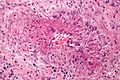
| Caption = Granulomatosis with polyangiitis. [[H&E stain]]. | |||
| Synonyms = Wegener's granulomatosis (old term) | |||
| Micro = | |||
| Subtypes = | |||
| LMDDx = small vessel vasculitis (inflammatory cells within the vessel wall, vessel wall injury - such as fibrinoid necrosis), granulomas - typically poorly formed | |||
| Stains = | |||
| IHC = | |||
| EM = | |||
| Molecular = | |||
| IF = | |||
| Gross = | |||
| Grossing = | |||
| Staging = | |||
| Site = [[blood vessels]] - see ''[[vasculitides]]'' | |||
| Assdx = renal failure (due to [[rapidly progressive glomerulonephritis]]), [[pulmonary hemorrhage]] | |||
| Syndromes = | |||
| Clinicalhx = | |||
| Signs = epistasis | |||
| Symptoms = | |||
| Prevalence = uncommon | |||
| Bloodwork = PR3-ANCA (c-ANCA) +ve | |||
| Rads = | |||
| Endoscopy = | |||
| Prognosis = | |||
| Other = | |||
| ClinDDx = dependent on presentation - nasal lesion: cocaine use; other causes of [[pulmonary hemorrhage]]; other causes of [[rapidly progressive glomerulonephritis]] | |||
| Tx = | |||
}} | |||
'''Granulomatosis with polyangiitis''', abbreviated '''GPA''', is a type of [[vasculitis]] that typically afflicts the [[lung]]s and [[kidney]]s. | |||
It was previously known as '''Wegener's granulomatosis''', abbreviated '''WG'''. | |||
It should '''''not''''' be confused with ''[[eosinophilic granulomatosis with polyangiitis]]'', previously known as ''Churg-Strauss syndrome''. | |||
==General== | ==General== | ||
| Line 6: | Line 42: | ||
===Clinical=== | ===Clinical=== | ||
*Epistasis. | *Epistasis. | ||
*Renal failure - | *Renal failure - presents as ''nephritic syndrome''. | ||
**Renal biopsy: crescentic glomerulonephritis ([[AKA]] [[rapidly progressive glomerulonephritis]]). | **Renal biopsy: crescentic glomerulonephritis ([[AKA]] [[rapidly progressive glomerulonephritis]]). | ||
*[[Pulmonary hemorrhage]]. | *[[Pulmonary hemorrhage]]. | ||
Serology: | Serology: | ||
*c-ANCA +ve.<ref name=Ref_TN2005_RH6>{{Ref TN2005|RH6}}</ref> | *PR3-ANCA (c-ANCA) +ve.<ref name=Ref_TN2005_RH6>{{Ref TN2005|RH6}}</ref> | ||
Notes: | Notes: | ||
Latest revision as of 22:01, 28 November 2016
Granulomatosis with polyangiitis, abbreviated GPA, is a type of vasculitis that typically afflicts the lungs and kidneys.
| Granulomatosis with polyangiitis | |
|---|---|
| Diagnosis in short | |
 Granulomatosis with polyangiitis. H&E stain. | |
|
| |
| Synonyms | Wegener's granulomatosis (old term) |
| LM DDx | small vessel vasculitis (inflammatory cells within the vessel wall, vessel wall injury - such as fibrinoid necrosis), granulomas - typically poorly formed |
| Site | blood vessels - see vasculitides |
|
| |
| Associated Dx | renal failure (due to rapidly progressive glomerulonephritis), pulmonary hemorrhage |
| Signs | epistasis |
| Prevalence | uncommon |
| Blood work | PR3-ANCA (c-ANCA) +ve |
| Clin. DDx | dependent on presentation - nasal lesion: cocaine use; other causes of pulmonary hemorrhage; other causes of rapidly progressive glomerulonephritis |
It was previously known as Wegener's granulomatosis, abbreviated WG.
It should not be confused with eosinophilic granulomatosis with polyangiitis, previously known as Churg-Strauss syndrome.
General
- Autoimmune.
Clinical
- Epistasis.
- Renal failure - presents as nephritic syndrome.
- Renal biopsy: crescentic glomerulonephritis (AKA rapidly progressive glomerulonephritis).
- Pulmonary hemorrhage.
Serology:
- PR3-ANCA (c-ANCA) +ve.[1]
Notes:
- Pulmonary hemorrhage syndromes:[2]
- Goodpasture syndrome.
- Idiopathic pulmonary hemosiderosis.
- Vasculitis-assoc. hemorrhage (hypersensitivity angiitis, Wegener granulomatosis).
- Systemic lupus erythematosus.
Microscopic
Features:
- Small vessel vasculitis:
- Inflammatory cells within the vessel wall.
- Granulomas - typically poorly formed.[3]
- Multinucleated giant cells - common. (???)
- Granulomas - typically poorly formed.[3]
- Vessel wall injury.
- Inflammatory cells within the vessel wall.
Notes:
- In the lung, the granulomas tend to be centrilobular, as the artery travels with the airway and is centrilobular.
- It may difficult to find small blood vessels in affected portions of lung.
Images
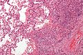
WG - intermed. mag. (WC)
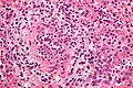
WG - very high mag. (WC)
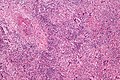
WG - intermed. mag. (WC)
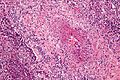
WG - high mag. (WC)
WG - very high mag. (WC)


www
See also
References
- ↑ Yeung, J.C.; Leonard, Blair J. N. (2005). The Toronto Notes 2005 - Review for the MCCQE and Comprehensive Medical Reference (2005 ed.). The Toronto Notes Inc. for Medical Students Inc.. pp. RH6. ISBN 978-0968592854.
- ↑ Cotran, Ramzi S.; Kumar, Vinay; Fausto, Nelson; Nelso Fausto; Robbins, Stanley L.; Abbas, Abul K. (2005). Robbins and Cotran pathologic basis of disease (7th ed.). St. Louis, Mo: Elsevier Saunders. pp. 745. ISBN 0-7216-0187-1.
- ↑ Cotran, Ramzi S.; Kumar, Vinay; Fausto, Nelson; Nelso Fausto; Robbins, Stanley L.; Abbas, Abul K. (2005). Robbins and Cotran pathologic basis of disease (7th ed.). St. Louis, Mo: Elsevier Saunders. pp. 747. ISBN 0-7216-0187-1.