Difference between revisions of "Gynecomastoid hyperplasia"
Jump to navigation
Jump to search
(split out) |
(more) |
||
| (4 intermediate revisions by the same user not shown) | |||
| Line 1: | Line 1: | ||
{{ Infobox diagnosis | |||
| Name = {{PAGENAME}} | |||
| Image = Gynecomastoid_hyperplasia_-_very_high_mag.jpg | |||
| Width = | |||
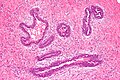
| Caption = Gynecomastoid hyperplasia. [[H&E stain]]. | |||
| Synonyms = gynecomastia | |||
| Micro = moderate hyperplasia - glands have more than 2 cell layers, "budding" (cells jut into the lumen, buds may be multicellular -- but narrower toward the centre of the lumen), stromal palor | |||
| Subtypes = | |||
| LMDDx = [[Micropapillary DCIS]] | |||
| Stains = | |||
| IHC = | |||
| EM = | |||
| Molecular = | |||
| IF = | |||
| Gross = | |||
| Grossing = | |||
| Site = [[breast]] | |||
| Assdx = [[Liver]] failure, [[Klinefelter syndrome]], testicular estrogen-producing [[germ cell tumour]] | |||
| Syndromes = | |||
| Clinicalhx = | |||
| Signs = excessive breast tissue | |||
| Symptoms = | |||
| Prevalence = | |||
| Bloodwork = | |||
| Rads = | |||
| Endoscopy = | |||
| Prognosis = benign | |||
| Other = | |||
| ClinDDx = | |||
| Tx = surgery | |||
}} | |||
'''Gynecomastoid hyperplasia''', also '''gynecomastia''', is a benign [[breast pathology|pathology of the breast]] classically seen in young men. | '''Gynecomastoid hyperplasia''', also '''gynecomastia''', is a benign [[breast pathology|pathology of the breast]] classically seen in young men. | ||
| Line 9: | Line 40: | ||
*[[Klinefelter syndrome]]. | *[[Klinefelter syndrome]]. | ||
*Testicular estrogen-producing [[germ cell tumour]]. | *Testicular estrogen-producing [[germ cell tumour]]. | ||
==Gross== | |||
*Excessive breast tissue in males. | |||
===Images=== | |||
<gallery> | |||
Image:Gynecomastia 001.jpg | Gynecomastia. (WC) | |||
Image:GynecomastiaFrontalAsymSevere.jpg | Gynecomastia - before and after. (WC) | |||
</gallery> | |||
==Microscopic== | ==Microscopic== | ||
| Line 30: | Line 70: | ||
*[http://www.hsc.stonybrook.edu/breast-atlas/XIII-03.htm Gynecomastoid hyperplasia (stonybrook.edu)]. | *[http://www.hsc.stonybrook.edu/breast-atlas/XIII-03.htm Gynecomastoid hyperplasia (stonybrook.edu)]. | ||
*[http://radiology.uchc.edu/eAtlas/Breast/1693.htm Gynecomastia (radiology.uchc.edu)]. | *[http://radiology.uchc.edu/eAtlas/Breast/1693.htm Gynecomastia (radiology.uchc.edu)]. | ||
==Sign out== | |||
<pre> | |||
A. Breast Tissue (60 g), Right, Excision: | |||
- Benign breast tissue. | |||
B. Breast Tissue (70 g), Left, Excision: | |||
- Benign breast tissue. | |||
</pre> | |||
===Alternate=== | |||
<pre> | |||
Left Chest Mass, Excision: | |||
- Breast tissue with gynecomastoid hyperplasia. | |||
- NEGATIVE for malignancy. | |||
</pre> | |||
===Micro=== | |||
The sections show breast tissue with epithelial hyperplasia and stromal palor. The architecture is normal. Epithelial budding is present. Significant atypia is absent. | |||
==See also== | ==See also== | ||
Latest revision as of 21:24, 16 November 2022
| Gynecomastoid hyperplasia | |
|---|---|
| Diagnosis in short | |
|
Template:Px Gynecomastoid hyperplasia. H&E stain. | |
|
| |
| Synonyms | gynecomastia |
|
| |
| LM | moderate hyperplasia - glands have more than 2 cell layers, "budding" (cells jut into the lumen, buds may be multicellular -- but narrower toward the centre of the lumen), stromal palor |
| LM DDx | Micropapillary DCIS |
| Site | breast |
|
| |
| Associated Dx | Liver failure, Klinefelter syndrome, testicular estrogen-producing germ cell tumour |
| Signs | excessive breast tissue |
| Prognosis | benign |
| Treatment | surgery |
Gynecomastoid hyperplasia, also gynecomastia, is a benign pathology of the breast classically seen in young men.
General
- Benign enlargement of breasts in males.
- Histologic changes may be seen in females.[1]
May be seen in the context of:
- Liver failure.
- Klinefelter syndrome.
- Testicular estrogen-producing germ cell tumour.
Gross
- Excessive breast tissue in males.
Images
- Gynecomastia 001.jpg
Gynecomastia. (WC)
- GynecomastiaFrontalAsymSevere.jpg
Gynecomastia - before and after. (WC)
Microscopic
Features:[1]
- Moderate hyperplasia.
- Glands have more than 2 cell layers.
- "Budding" - individual cells jut into the lumen - key feature.
- Buds may be multicellular; however, narrower toward the centre of the lumen.
- Stromal palor.[2]
DDx:
- Micropapillary DCIS - buds not narrower toward the centre of the lumen.
Images
Gynecomastoid hyperplasia - intermed. mag. (WC/Nephron)
Gynecomastoid hyperplasia - very high mag. (WC/Nephron)
Gynecomastoid hyperplasia - 2 - intermed. mag. (WC/Nephron)

www:
Sign out
A. Breast Tissue (60 g), Right, Excision:
- Benign breast tissue.

B. Breast Tissue (70 g), Left, Excision:
- Benign breast tissue.
Alternate
Left Chest Mass, Excision:
- Breast tissue with gynecomastoid hyperplasia.
- NEGATIVE for malignancy.
Micro
The sections show breast tissue with epithelial hyperplasia and stromal palor. The architecture is normal. Epithelial budding is present. Significant atypia is absent.
See also
References
- ↑ 1.0 1.1 URL: http://www.hsc.stonybrook.edu/breast-atlas/XIII-03.htm. Accessed on: 16 November 2011.
- ↑ URL: http://radiology.uchc.edu/eAtlas/Breast/1693.htm. Accessed on: 16 November 2011.