Difference between revisions of "Columnar cell change of the breast"
Jump to navigation
Jump to search

| (One intermediate revision by the same user not shown) | |||
| Line 1: | Line 1: | ||
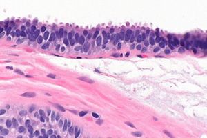
[[Image:Columnar cell change -- very high mag.jpg |thumb|right|Columnar cell change of the breast - very high magnification. [[H&E stain]]. (WC/Nephron)]] | |||
'''Columnar cell change of the breast''', usually '''columnar cell change''' (abbreviated '''CCC'''), is a benign finding in [[breast pathology]]. | '''Columnar cell change of the breast''', usually '''columnar cell change''' (abbreviated '''CCC'''), is a benign finding in [[breast pathology]]. | ||
| Line 13: | Line 14: | ||
**The snouts are attached to the cell-- appear as round ball only in the plane of section. | **The snouts are attached to the cell-- appear as round ball only in the plane of section. | ||
*Cytoplasm +/-eosinophilia. | *Cytoplasm +/-eosinophilia. | ||
*Often purple luminal calcifications | *Often (purple) luminal [[breast calcifications|calcifications]]. | ||
DDx: | DDx: | ||
*Flat epithelial atypia (>2 cell layers).{{Fact}} | *[[Flat epithelial atypia]] (>2 cell layers).{{Fact}} | ||
**If the columnar cells shows low to intermediate grade atypia the process is termed "flat epithelial atypia" | **If the columnar cells shows low to intermediate grade atypia the process is termed "flat epithelial atypia" | ||
**If higher grade | **If higher grade atypia is present the lesion is termed "flat DCIS" (clinging carcinoma). | ||
===Images=== | ===Images=== | ||
Latest revision as of 21:08, 26 November 2021

Columnar cell change of the breast - very high magnification. H&E stain. (WC/Nephron)
Columnar cell change of the breast, usually columnar cell change (abbreviated CCC), is a benign finding in breast pathology.
It is also known as blunt duct adenosis.
General
- Columnar cell change is associated with (benign) calcification[1] - key point.
Microscopic
Features:
- Secretory cells (line gland lumen) have columnar morphology.
- May have "apical snouts".
- Blebs or round balls eosinophilic material appear to be adjacent to the cell at their luminal surface.
- The snouts are attached to the cell-- appear as round ball only in the plane of section.
- Cytoplasm +/-eosinophilia.
- Often (purple) luminal calcifications.
DDx:
- Flat epithelial atypia (>2 cell layers).[citation needed]
- If the columnar cells shows low to intermediate grade atypia the process is termed "flat epithelial atypia"
- If higher grade atypia is present the lesion is termed "flat DCIS" (clinging carcinoma).
Images
www=
Sign out
- Usually not reported.
See also
References
- ↑ Jara-Lazaro, AR.; Tse, GM.; Tan, PH. (Jan 2009). "Columnar cell lesions of the breast: an update and significance on core biopsy.". Pathology 41 (1): 18-27. doi:10.1080/00313020802563486. PMID 19089736.


