Difference between revisions of "Ganglioglioma"
Jump to navigation
Jump to search
Jensflorian (talk | contribs) (→Molecular: update) |
Jensflorian (talk | contribs) (→DDx:: DIA/DIG added) |
||
| Line 119: | Line 119: | ||
*[[Oligodendroglioma]]. | *[[Oligodendroglioma]]. | ||
*[[PXA]]. | *[[PXA]]. | ||
*Desmoplastic infantile astrocytoma and ganglioglioma. | |||
*Cortical tuber. | *Cortical tuber. | ||
*Trapped cortical neurons in diffuse astrocytoma. | *Trapped cortical neurons in diffuse astrocytoma. | ||
Revision as of 10:50, 14 September 2017
| Ganglioglioma | |
|---|---|
| Diagnosis in short | |
 | |
| LM DDx | piloid gliosis, pilocytic astrocytoma, DNT |
| Stains | PAS-D +ve (eosinophilic granular bodies) |
| IHC | GFAP +ve, Synapto +ve |
| Gross | usually temporal +/-cystic |
| Site | brain - usu. supratentorial |
|
| |
| Syndromes | associated with epilepsy |
|
| |
| Prevalence | rare - esp. in children |
| Prognosis | good (WHO Grade I) |
Ganglioglioma is a epilepsy-associated glioneuronal tumour with benign course. Not to be confused with ganglioneuroma.
General
- Gangliolioma: Grade I WHO mixed neuronal-glial tumour (ICD-O code: 9505/1).
- Anaplastic ganglioglioma: Grade III (ICD-O: 9505/3)
- Rare (approx. 0.5% of all CNS tumors).
- Usu. temporal lobe.
- Predominantly children (mean age: 9 years).
- Recognized as a cause of epilepsy.[1]
- Favourable prognosis (survival rates up to 97%)
- Anaplastic ganglioglioma have a recurrence risk of 69%[2]
Imaging
- Well-defined, T2-hyperintense.
- Strong CM enhancement.
- May contain cysts.
- Associated with temporal lobe.
Gross
- Circumscribed lesion.
- Usu. contrast enhancing.
- Solid, but intracortical cysts may be present.
- Little mass effect.
Microscopic
Microscopic
Features:
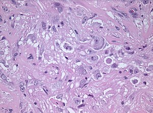
- Dysplastic neurons.
- Out of regular architecture / abnormal location.
- Cytomegaly
- Clustering
- Binucleated (very occassionally).
- Atypical glia (ie neoplastic).
- Eosinophilic granular bodies (more common than rosenthal fibers).
- Dystrophic calcification.
- Prominent capillary network.
- Lymphocytic cuffing.
- May contain some reticulin.
- Glial component may resemble:
- Fibrillary astrocytoma.
- Oligodendroglioma.
- Pilocytic astrocytoma.
Anaplastic ganglioglioma:
- Brisk mitotic activity
- Necrosis
IHC
- Neurons:
- MAP2 +ve
- Synaptophysin +ve
- Neurofilament +ve
- Chromogranin +ve
- Glia:
- CD34+/-ve
- BRAF V600E +ve (approx. 25%, mainly ganglion cells).
- MAP2: usu. absent.
- MIB-1 (low, but resembles proliferative tumor component).
Molecular
- BRAF V600E-mutated(approx. 25%).[3]
- BRAF V600E antibody stains especially neuronal cells.[4]
- IDH1/2 wt.
- No 1p/19q codeletion.
- Usu. Chr. 7 gain.
- Rare cases with KIAA1459-BRAF fusion.[5]
- CDKN2A deletions in anaplastic ganglioglioma.
- rare cases with co-occurrence of K27M mutation.[6]
Images
Prognosis
- Very good (10-year OS: 97%)
- Primary treatment: surgery.
- Seizure free outcome: 81%.
- Incomplete resection as major factor for persisting epilepsia.[7]
DDx:
- DNT.
- Oligodendroglioma.
- PXA.
- Desmoplastic infantile astrocytoma and ganglioglioma.
- Cortical tuber.
- Trapped cortical neurons in diffuse astrocytoma.
- Papillary glioneuronal tumor.
See also
References
- ↑ Im, SH.; Chung, CK.; Cho, BK.; Lee, SK. (Mar 2002). "Supratentorial ganglioglioma and epilepsy: postoperative seizure outcome.". J Neurooncol 57 (1): 59-66. PMID 12125968.
- ↑ Terrier, LM.; Bauchet, L.; Rigau, V.; Amelot, A.; Zouaoui, S.; Filipiak, I.; Caille, A.; Almairac, F. et al. (05 2017). "Natural course and prognosis of anaplastic gangliogliomas: a multicenter retrospective study of 43 cases from the French Brain Tumor Database.". Neuro Oncol 19 (5): 678-688. doi:10.1093/neuonc/now186. PMID 28453747.
- ↑ Schindler, G.; Capper, D.; Meyer, J.; Janzarik, W.; Omran, H.; Herold-Mende, C.; Schmieder, K.; Wesseling, P. et al. (Mar 2011). "Analysis of BRAF V600E mutation in 1,320 nervous system tumors reveals high mutation frequencies in pleomorphic xanthoastrocytoma, ganglioglioma and extra-cerebellar pilocytic astrocytoma.". Acta Neuropathol 121 (3): 397-405. doi:10.1007/s00401-011-0802-6. PMID 21274720.
- ↑ Koelsche, C.; Wöhrer, A.; Jeibmann, A.; Schittenhelm, J.; Schindler, G.; Preusser, M.; Lasitschka, F.; von Deimling, A. et al. (Jun 2013). "Mutant BRAF V600E protein in ganglioglioma is predominantly expressed by neuronal tumor cells.". Acta Neuropathol 125 (6): 891-900. doi:10.1007/s00401-013-1100-2. PMID 23435618.
- ↑ Mesturoux, L.; Durand, K.; Pommepuy, I.; Robert, S.; Caire, F.; Labrousse, F. (Aug 2016). "Molecular Analysis of Tumor Cell Components in Pilocytic Astrocytomas, Gangliogliomas, and Oligodendrogliomas.". Appl Immunohistochem Mol Morphol 24 (7): 496-500. doi:10.1097/PAI.0000000000000288. PMID 27389560.
- ↑ Pagès, M.; Beccaria, K.; Boddaert, N.; Saffroy, R.; Besnard, A.; Castel, D.; Fina, F.; Barets, D. et al. (Dec 2016). "Co-occurrence of histone H3 K27M and BRAF V600E mutations in paediatric midline grade I ganglioglioma.". Brain Pathol. doi:10.1111/bpa.12473. PMID 27984673.
- ↑ Devaux, B.; Chassoux, F.; Landré, E.; Turak, B.; Laurent, A.; Zanello, M.; Mellerio, C.; Varlet, P. (Jun 2017). "Surgery for dysembryoplastic neuroepithelial tumors and gangliogliomas in eloquent areas. Functional results and seizure control.". Neurochirurgie 63 (3): 227-234. doi:10.1016/j.neuchi.2016.10.009. PMID 28506485.


