Difference between revisions of "Angiosarcoma"
Jump to navigation
Jump to search
(split-out) |
|||
| Line 12: | Line 12: | ||
==Microscopic== | ==Microscopic== | ||
Features: | Features: | ||
*Spindle cell lesion. | |||
**Occasionally an epithelioid lesion. | |||
*Very many small capillaries of irregular shape lined with: | *Very many small capillaries of irregular shape lined with: | ||
**Pleomorphic nuclei. | **Pleomorphic nuclei - '''important'''. | ||
***May have hobnail morphology. | ***May have hobnail morphology. | ||
**Usually "red" at low power - due to many RBCs - '''important'''. | |||
*Mitoses. | *Mitoses. | ||
*Cytoplasmic vacuoles. | *Cytoplasmic vacuoles. | ||
Revision as of 20:16, 9 October 2013
Angiosarcoma is an uncommon malignant vascular tumour.
General
- Malignant tumour - general has a poor prognosis.[1]
Epidemiology:
- May arise secondary to chronic lymphedema related to breast carcinoma.
- Known as Stewart–Treves syndrome.[2]
- Liver angiosarcomas are associated with vinyl chloride exposure.[3]
- Cutaneous angiosarcomas are classically seen on the head and neck of whites over 60 years old.[4]
Microscopic
Features:
- Spindle cell lesion.
- Occasionally an epithelioid lesion.
- Very many small capillaries of irregular shape lined with:
- Pleomorphic nuclei - important.
- May have hobnail morphology.
- Usually "red" at low power - due to many RBCs - important.
- Pleomorphic nuclei - important.
- Mitoses.
- Cytoplasmic vacuoles.
- Cells trying to form lumina - embryologic.
Notes:
- Epithelioid variant (with abundant cytoplasm & sheeting architecture) may resemble melanoma or hepatocellular carcinoma.
DDx:
- Atypical vascular lesion.
- Kaposi sarcoma.
- Poorly differentiated carcinoma.
Images
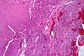
Epithelioid angiosarcoma - very low mag. (WC/Nephron)
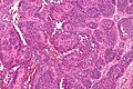
Epithelioid angiosarcoma - intermed mag. (WC/Nephron)
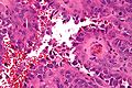
Epithelioid angiosarcoma - very high mag. (WC/Nephron)
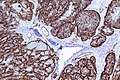
Epithelioid angiosarcoma - CD31 - intermed. mag. (WC/Nephron)


IHC
See also
References
- ↑ Young RJ, Brown NJ, Reed MW, Hughes D, Woll PJ (May 2010). "Angiosarcoma". Lancet Oncol. doi:10.1016/S1470-2045(10)70023-1. PMID 20537949.
- ↑ Pincus, LB.; Fox, LP. (Aug 2008). "Images in clinical medicine. The Stewart-Treves syndrome.". N Engl J Med 359 (9): 950. doi:10.1056/NEJMicm071344. PMID 18753651. http://www.nejm.org/doi/full/10.1056/NEJMicm071344.
- ↑ Mitchell, Richard; Kumar, Vinay; Fausto, Nelson; Abbas, Abul K.; Aster, Jon (2011). Pocket Companion to Robbins & Cotran Pathologic Basis of Disease (8th ed.). Elsevier Saunders. pp. 212. ISBN 978-1416054542.
- ↑ Albores-Saavedra, J.; Schwartz, AM.; Henson, DE.; Kostun, L.; Hart, A.; Angeles-Albores, D.; Chablé-Montero, F. (Apr 2011). "Cutaneous angiosarcoma. Analysis of 434 cases from the Surveillance, Epidemiology, and End Results Program, 1973-2007.". Ann Diagn Pathol 15 (2): 93-7. doi:10.1016/j.anndiagpath.2010.07.012. PMID 21190880.
- ↑ Rossi, S.; Orvieto, E.; Furlanetto, A.; Laurino, L.; Ninfo, V.; Dei Tos, AP. (May 2004). "Utility of the immunohistochemical detection of FLI-1 expression in round cell and vascular neoplasm using a monoclonal antibody.". Mod Pathol 17 (5): 547-52. doi:10.1038/modpathol.3800065. PMID 15001993.
- ↑ Kahn, HJ.; Bailey, D.; Marks, A. (Apr 2002). "Monoclonal antibody D2-40, a new marker of lymphatic endothelium, reacts with Kaposi's sarcoma and a subset of angiosarcomas.". Mod Pathol 15 (4): 434-40. doi:10.1038/modpathol.3880543. PMID 11950918.