Spindle cell
Jump to navigation
Jump to search
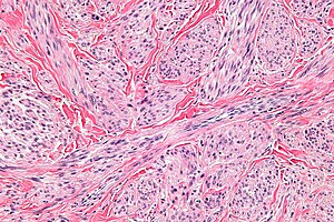


Spindle cells in a leiomyosarcoma. (WC)
Spindle cell is a histomorphologic descriptor used in pathology.
A list of spindle cell lesions is found the in the spindle cell lesions article.
Definition
It refers to a cell that is tapered at both ends.[1]
Notes:
- A taper gradually decreases toward one end [of the cross-section or width].[2]
- Image: Taperred thread (qcfocus.com).
- Spindle cells can have "pointy" ends (typical for nerves) or "rounded" ends (typical for muscle), i.e. be ellipitcal or vesica piscis.
Subtyping spindle cells by H&E
Spindle cells can often be subtyped based on H&E:[3]
- Fibroblast = blue.
- Smooth muscle = deep pink.
- Myofibroblast = purple.
Images
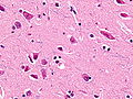
Spindle neurons. (WC)
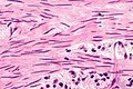
Benign smooth muscle cells of the urinary bladder. (WC)
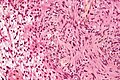
Spindle cells of a schwannoma. (WC)


Shapes

A spindle. (WC)

Vesica piscis. (WC)
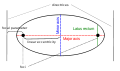
Ellipse. (WC)


See also
References
- ↑ URL: http://www.medterms.com/script/main/art.asp?articlekey=25657. Accessed on: 2 February 2011.
- ↑ URL: http://dictionary.reference.com/browse/taper. Accessed on: 3 February 2011.
- ↑ Chan, JK. (Feb 2014). "The wonderful colors of the hematoxylin-eosin stain in diagnostic surgical pathology.". Int J Surg Pathol 22 (1): 12-32. doi:10.1177/1066896913517939. PMID 24406626.