Pseudopyloric mucous glands
Jump to navigation
Jump to search


Pseudopyloric mucous glands, also pyloric gland metaplasia,[1] is a change seen the intestine. It is associated with Crohn's disease.
Pseudopyloric mucous glands is abbreviated PMG. Pyloric gland metaplasia is abbreviated PGM.
General
- May be seen in Crohn's disease in the terminal ileum.[2]
- Suggestive of Crohn's disease in pouchitis but not diagnostic.[3]
Microscopic
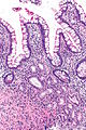
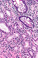
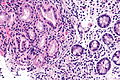
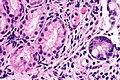
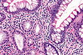
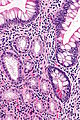
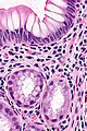
Features:[4]
- Round glands composed of tall columnar cells with:
- Abundant pale (pink) cytoplasm.
- Small basal nuclei.
- Usually in the deep aspect of the mucosa.
Note:
- The lining cells look somewhat like stubby champagne flutes.
Images

PMG - low mag.
PMG - intermed. mag.
PMG - high mag.
PMG - high mag.
PMG - very high mag.


PMG - high mag.
PMG - high mag.
PMG - very high mag.


Champagne flute. (WC)

See also
References
- ↑ URL: http://www.medunigraz.at/22698. Accessed on: 6 August 2013.
- ↑ Geboes, K.; Ectors, N.; D'Haens, G.; Rutgeerts, P. (Feb 1998). "Is ileoscopy with biopsy worthwhile in patients presenting with symptoms of inflammatory bowel disease?". Am J Gastroenterol 93 (2): 201-6. doi:10.1111/j.1572-0241.1998.00201.x. PMID 9468242.
- ↑ Agarwal, S.; Stucchi, AF.; Dendrinos, K.; Cerda, S.; O'Brien, MJ.; Becker, JM.; Heeren, T.; Farraye, FA. (Oct 2013). "Is pyloric gland metaplasia in ileal pouch biopsies a marker for Crohn's disease?". Dig Dis Sci 58 (10): 2918-25. doi:10.1007/s10620-013-2655-4. PMID 23543088.
- ↑ Weber, CR.; Rubin, DT. (Oct 2013). "Chronic pouchitis versus recurrent Crohn's disease: a diagnostic challenge.". Dig Dis Sci 58 (10): 2748-50. doi:10.1007/s10620-013-2816-5. PMID 23925821.