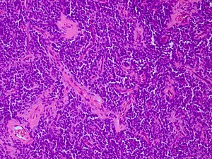
Other CNS embryonal tumours
Jump to navigation
Jump to search
| Other CNS embryonal tumours | |
|---|---|
| Diagnosis in short | |
 Other CNS embryonal tumorH&E stain. | |
|
| |
| Synonyms | CNS-PNET |
| LM DDx | small round blue cell tumours |
| IHC | S-100 +ve, Syn +/-ve |
| Site | brain, spinal cord |
|
| |
| Prevalence | rare - typically in children and young adults |
| Prognosis | poor (WHO Grade IV) |
Other CNS embryonal tumor, was introduced in the WHO 2016 classification of CNS tumors. Some of these tumors of this category has been previously designated as CNS-PNET but this terminology has been abandoned.
General
Other CNS embryonal tumours are a group of primitive neuroeptithelial tumors that are to be classified in distinct molecular groups in the future. Currently it is a exlcusion criteria encompassing four morphological subgroups
- CNS neuroblastomas
- often infants.
- Tumors can be huge.
- CNS ganglioneuroblastomas.
- often characterized by FOXR2 fusions: CNS neuroblastoma with FOXR2 activation (NB‐FOXR2)
- CNS medulloepithelioma.
- but absence of C19MC alteration, otherwise classified as ETMR
- CNS embryonal tumour, NOS
- Similarities to astroblastoma: High‐grade neuroepithelial tumours with MN1 alteration (HGNET‐MN1)
- NUTM1-positive IHC: Ewing's sarcoma family tumour with CIC alteration (EFT‐CIC)
- BCOR-positive IHC: HGNET BCOR‐altered neuroepithelial tumours
Currently four distinct subtypes can be discriminated by molecular analysis: NB-FOXR2, HGNET-MN1, EFT-CIC and HGNET-BCOR.[1]
Histology
CNS neuroblastoma:
- Groups of neurocytic cells.
- Variable neuropil-rich stroma.
- Small round blue cells.
HGNET-BCOR:
- Circumscribed growth.
- Perivascular pesudorosettes.
- Fibrillary areas resembling glioma.
- Palisading necrosis.
- Absence of microvascular proliferation.
IHC
CNS Ganglioneuroblastoma + Neuroblastoma
- Syn -/+ve.
- Vim -ve.
- GFAP usu -ve.
- MIB-1 usu high.
Medulloepthelioma
- Syn +/-ve.
- NF +/-ve.
- GFAP usu -ve.
- CK may be focally +ve.
HGNET-BCOR [2]
- OLIG variable +ve.
- BCOR +ve.
- Syn -ve.
- GFAP -ve.
- EMA: dots +ve.
- NEUN +/-ve.
DDx:
- AT/RT
- ETMR
- Anaplastic ependymoma
- H3F3A G34-mutated Glioblastoma
- Metastasis of Ewing sarcoma
- ↑ Sturm D, Orr BA, Toprak UH, Hovestadt V, Jones DTW, Capper D, Sill M, Buchhalter I, Northcott PA, Leis I, Ryzhova M, Koelsche C, Pfaff E, Allen SJ, Balasubramanian G, Worst BC, Pajtler KW, Brabetz S, Johann PD, Sahm F, Reimand J, Mackay A, Carvalho DM, Remke M, Phillips JJ, Perry A, Cowdrey C, Drissi R, Fouladi M, Giangaspero F, Łastowska M, Grajkowska W, Scheurlen W, Pietsch T, Hagel C, Gojo J, Lötsch D, Berger W, Slavc I, Haberler C, Jouvet A, Holm S, Hofer S, Prinz M, Keohane C, Fried I, Mawrin C, Scheie D, Mobley BC, Schniederjan MJ, Santi M, Buccoliero AM, Dahiya S, Kramm CM, von Bueren AO, von Hoff K, Rutkowski S, Herold-Mende C, Frühwald MC, Milde T, Hasselblatt M, Wesseling P, Rößler J, Schüller U, Ebinger M, Schittenhelm J, Frank S, Grobholz R, Vajtai I, Hans V, Schneppenheim R, Zitterbart K, Collins VP, Aronica E, Varlet P, Puget S, Dufour C, Grill J, Figarella-Branger D, Wolter M, Schuhmann MU, Shalaby T, Grotzer M, van Meter T, Monoranu CM, Felsberg J, Reifenberger G, Snuderl M, Forrester LA, Koster J, Versteeg R, Volckmann R, van Sluis P, Wolf S, Mikkelsen T, Gajjar A, Aldape K, Moore AS, Taylor MD, Jones C, Jabado N, Karajannis MA, Eils R, Schlesner M, Lichter P, von Deimling A, Pfister SM, Ellison DW, Korshunov A, Kool M (February 2016). "New Brain Tumor Entities Emerge from Molecular Classification of CNS-PNETs". Cell 164 (5): 1060–1072. doi:10.1016/j.cell.2016.01.015. PMC 5139621. PMID 26919435. https://www.ncbi.nlm.nih.gov/pmc/articles/PMC5139621/.
- ↑ Ferris SP, Velazquez Vega J, Aboian M, Lee JC, Van Ziffle J, Onodera C, Grenert JP, Saunders T, Chen YY, Banerjee A, Kline CN, Gupta N, Raffel C, Samuel D, Ruiz-Diaz I, Magaki S, Wilson D, Neltner J, Al-Hajri Z, Phillips JJ, Pekmezci M, Bollen AW, Tihan T, Schniederjan M, Cha S, Perry A, Solomon DA (January 2020). "High-grade neuroepithelial tumor with BCOR exon 15 internal tandem duplication-a comprehensive clinical, radiographic, pathologic, and genomic analysis". Brain Pathol. 30 (1): 46–62. doi:10.1111/bpa.12747. PMID 31104347.