Synovial chondromatosis
(Redirected from Loose bodies)
Jump to navigation
Jump to search
| Synovial chondromatosis | |
|---|---|
| Diagnosis in short | |
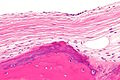
|
Template:Px Loose body. H&E stain. | |
|
| |
| LM | hyaline cartilage +/- lobular surface +/-lacunae with binucleate cells, +/-nuclear atypia (moderate to severe), +/-synovial hyperplasia, bone |
| LM DDx | chondrosarcoma, osteoarthritis |
| Site | joints - classically knee or hip |
|
| |
| Symptoms | pain |
| Prognosis | benign |
| Treatment | removal of loose bodies |
Synovial chondromatosis is a relative common pathology of the joint. It is also known as synovial osteochondromatosis.
Loose body, loose bodies, rice bodies and joint mice redirect to this page.
General
- Benign.
- Malignant transformation rare <5%.[1]
- Classically location: knee.[1]
- Hip next most common site.
- Usually adults.
- Prevalence: male > female.
Note:
- This is a clinicoradiologic diagnosis.
Gross/radiology
- Intraarticular calcifications.
- Diffuse involvement of the joint.
- +/-Loose bodies in the joint (AKA joint mice).
Image
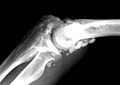
Synovial chondromatosis. (WC)

www:
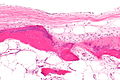
Microscopic
Features:[1]
- Hyaline cartilage +/- lobular surface.
- +/-Lacunae with binucleate cells.
- +/-Nuclear atypia - moderate to severe.[2]
- +/-Synovial hyperplasia - ribbon like tissue with an epithelium that has eosinophilic cytoplasm.
- Bone.
DDx:
Images
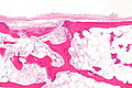
LB - low mag.
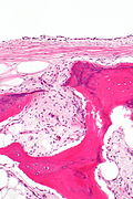
LB - intermed. mag.

LB - high mag.

LB - very high mag.
LB - intermed. mag.
LB - high mag.

LB - very high mag.


www:
- Synovial chondromatosis (rsna.org).
- Synovial chondromatosis - low mag. (rsna.org).
- Synovial chondromatosis (webpathology.com).
- Loose body with fibrotic synovial membrane (nih.gov).
Sign out
Synovium, Left Knee, Biopsy:
     - Synovium with mild chronic inflammation.
     - Cartilage with ossification, compatible with loose bodies/rice bodies.
     - NEGATIVE for evidence of malignancy.
Block letters
LOOSE BODIES, RIGHT ELBOW, REMOVAL:
- FRAGMENTS OF BONE WITH CARTILAGE AND SYNOVIAL TISSUE COMPATIBLE WITH LOOSE BODIES.
TISSUE, CAPSULE LEFT ELBOW, REMOVAL:
- DEGENERATIVE JOINT DISEASE.
- SYNOVIAL HYPERPLASIA WITHOUT SIGNIFICANT INFLAMMATION.
- ROUND BODIES CONSISTING OF BENIGN BONE WITH A FATTY MARROW, AND FIBROUS
  AND CARTILAGINOUS SURFACE, COMPATIBLE WITH LOOSE BODIES.
- NEGATIVE FOR MALIGNANCY.
LOOSE BODY, LEFT KNEE, REMOVAL:
- BENIGN BONE WITH FIBROTIC SYNOVIAL MEMBRANE, CONSISTENT WITH LOOSE BODY.
Micro
The sections show multiple fragments of tissue consisting of bone covered by hyaline cartilage and associated with synovial hyperplasia.
There is no appreciable nuclear atypia or mitotic activity.
See also
References
- ↑ 1.0 1.1 1.2 Murphey, MD.; Vidal, JA.; Fanburg-Smith, JC.; Gajewski, DA.. "Imaging of synovial chondromatosis with radiologic-pathologic correlation.". Radiographics 27 (5): 1465-88. doi:10.1148/rg.275075116. PMID 17848703.
- ↑ URL: http://www.webpathology.com/image.asp?n=3&Case=369. Accessed on: 10 December 2012.