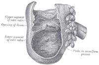
Ileocecal valve
Jump to navigation
Jump to search
The ileocecal valve, abbreviated IC valve, is the divider between the small bowel and cecum. It is seen by pathologist in some subtotal colectomies (e.g. right hemicoloectomies) and occasionally biopsied.
Lipomatous ileocecal valve
- AKA lipomatosis of the ileocecal valve
General
- The lesion should involve the valve circumferentially.
- True lipomas of the ileocecal have a capsule, are not circumferential and less common.[1]
Clinical:
- May be misdiagnosed as malignancy.[2]
- Reported to mimic Crohn's disease.[3]
Gross
- "Ileocecal valve prominent".
Microscopic
Feature:
- Mature adipocytes.
- No capsule.[1]
DDx:
- Lipoma of the ileocecal valve - have a capsule.
Image:
Sign out
ILEOCECAL VALVE, BIOPSY:
- SUBMUCOSA WITH A LARGE CLUSTER OF MATURE ADIPOCYTES, SEE COMMENT.
- BOWEL MUCOSA WITHIN NORMAL LIMITS.

COMMENT:
The findings are consistent with a lipomatous ileocecal valve.
Small amount of adipose tissue
ILEOCECAL VALVE ("PROMINENT"), BIOPSY:
- COLONIC-TYPE MUCOSA WITH PROMINENT PANETH CELLS AND FOCAL LAMIMA
  PROPRIA NEUTROPHILS.
- SMALL AMOUNT OF BENIGN (SUBMUCOSAL) ADIPOSE TISSUE.
- NO DEFINITE ACUTE VALVITIS.
- NEGATIVE FOR DYSPLASIA.
No submucosa
ILEOCECAL VALVE, BIOPSY:
- COLONIC-TYPE MUCOSA WITHIN NORMAL LIMITS.
- NO SUBMUCOSA PRESENT.
Ileocecal tuberculosis
General
- Ileocecal region and jejunoileal region are the most commonly affected areas in gastrointestinal tuberculosis.[5][6][7]
Microscopic
- See Tuberculosis.
Ileocecal valve ulceration
General
- Relatively uncommon.
Microscopic
Features:
- Fibrin - acellular amorphous eosinophilic material.
- Neutrophils.
- Cryptitis (focal).
DDx:
- Crohn's disease.
- NSAID-associated ulcer.[8]
- Infection, e.g. tuberculosis.
- Idiopathic - common for small lesions.[8]
- Mechanical forces (shear) - in a prominent IC valve.
- Shearing forces are certainly present[9]... not studied.
Sign out
Early changes due to mechanical factors in a prominent valve
ILEOCECAL VALVE, BIOPSY:
- SMALL BOWEL MUCOSA WITH FOCAL CRYPTITIS, SEE COMMENT.
-- NEGATIVE FOR GRANULOMAS AND NEGATIVE FOR ARCHITECTURAL DISTORTION.
-- NEGATIVE FOR DYSPLASIA.

COMMENT:
The clinical history is noted. No adipose tissue is seen in this superficial mucosal
biopsy. The cryptitis is seen focally at the tips of well-formed villi. This could be due
to mechanical factors; however, other causes should be considered clinically.
ILEOCEAL VALVE, BIOPSY:
- BOWEL MUCOSA WITH MILD ACTIVE INFLAMMATION, SEE COMMENT.
-- NEGATIVE FOR GRANULOMAS.
-- NEGATIVE FOR DYSPLASIA.

COMMENT:
The clinical history (prominent ileocecal valve) is noted. No adipose tissue is seen in
this superficial mucosal biopsy.

The inflammation is focal and superficial, and blunted-appearing villi are present. The
changes may be due to mechanical factors; however, other causes should be considered
clinically.
See also
References
- ↑ 1.0 1.1 Skaane, P.; Eide, TJ.; Westgaard, T.; Gauperaa, T. (Dec 1981). "Lipomatosis and true lipomas of the ileocecal valve.". Rofo 135 (6): 663-8. doi:10.1055/s-2008-1056492. PMID 6212382.
- ↑ Petrović, J.; Barisić, G.; Saranović, D.; Micev, M.; Krivokapić, Z. (Sep 2007). "Lipomatosis of the ileocecal valve treated with right hemicolectomy as the consequence of an incomplete diagnostic procedure.". Tech Coloproctol 11 (3): 278-80. doi:10.1007/s10151-007-0366-6. PMID 17676259.
- ↑ Bhupalan, AJ.; Forbes, A.; Lloyd-Davies, E.; Wignall, B.; Murray-Lyon, IM. (Jun 1992). "Lipomatosis of the ileocaecal valve simulating Crohn's disease.". Postgrad Med J 68 (800): 455-6. PMID 1437927.
- ↑ Dultz, LA.; Ullery, BW.; Sun, HH.; Huston, TL.; Eachempati, SR.; Barie, PS.; Shou, J.. "Ileocecal valve lipoma with refractory hemorrhage.". JSLS 13 (1): 80-3. PMC 3015905. PMID 19366548. https://www.ncbi.nlm.nih.gov/pmc/articles/PMC3015905/.
- ↑ Iacobuzio-Donahue, Christine A.; Montgomery, Elizabeth A. (2005). Gastrointestinal and Liver Pathology: A Volume in the Foundations in Diagnostic Pathology Series (1st ed.). Churchill Livingstone. pp. 282. ISBN 978-0443066573.
- ↑ Bhargava, DK.; Tandon, HD.; Chawla, TC.; Shriniwas, BN.; Tandon, BM.; Kapur, . (Apr 1985). "Diagnosis of ileocecal and colonic tuberculosis by colonoscopy.". Gastrointest Endosc 31 (2): 68-70. PMID 3922847.
- ↑ Engin, G.; Balk, E.. "Imaging findings of intestinal tuberculosis.". J Comput Assist Tomogr 29 (1): 37-41. PMID 15665681.
- ↑ 8.0 8.1 Liu, JX.; Wang, HH. (Mar 2008). "[Clinical and pathological features of benign ileocecal ulcerative lesions discovered by ileocolonoscopy: analysis of 31 cases].". Zhonghua Yi Xue Za Zhi 88 (12): 823-5. PMID 18756986.
- ↑ Gayer, CP.; Basson, MD. (Aug 2009). "The effects of mechanical forces on intestinal physiology and pathology.". Cell Signal 21 (8): 1237-44. doi:10.1016/j.cellsig.2009.02.011. PMID 19249356.
