Gallbladder carcinoma
| Gallbladder carcinoma | |
|---|---|
| Diagnosis in short | |
_histopathology.jpg) Gallbladder adenocarcinoma. H&E stain. | |
|
| |
| LM | atypical epithelium usually gland forming, may be bland with an infiltrative growth pattern |
| LM DDx | adenomyoma of the gallbladder, metastatic carcinoma (e.g. cholangiocarcinoma), gallbladder adenoma |
| IHC | CK7 +ve, CK20 -ve, CDX2 -ve |
| Gross | lesion - usually fundus of gallbladder |
| Grossing notes | gallbladder grossing |
| Site | gallbladder |
|
| |
| Associated Dx | chronic cholecystitis, gallstones, primary sclerosing cholangitis, intestinal metaplasia of the gallbladder |
| Prevalence | uncommon |
Gallbladder carcinoma is a malignant epithelial neoplasm arising from the gallbladder. Most gallbladder carcinomas are adenocarcinomas.
General
- Uncommon.
Treatment:
- Cholecystectomy +/- lymph nodes +/- partial hepatectomy.[1]
Epidemiology
- Associated with gallstones.
- Increased risk in primary sclerosing cholangitis.
- Sex: female > male.
- Location: usually fundus, sometimes body.
Notes:
- Diffuse calcification of gallbladder wall, AKA "porcelain gallbladder" is not associated with carcinoma - based on a series of 10,741 cholecystectomies.[2]
- Focal mucosal calcification is associated with malignancy.[3]
- Cholangiocarcinoma is dealt with in the liver neoplasms article.
Gross
- Classic: mass projecting into the lumen.
- Marked gallbladder wall thickening.
- >10 mm should be considered with suspicion.[4]
Image:
Microscopic
Features:
- Usually adenocarcinoma.
- Mimics appearance of pancreatic ductal adenocarcinoma -- but less cellular mucin.[5]
Notes:
- May be very subtle, i.e. difficult to differentiate from normal glands.
- "Deep glands" that look bland shouldn't immediately be dismissed as benign.
Subtypes:
DDx:
- Adenomyoma of the gallbladder.
- Gallbladder adenoma.
- Metastatic carcinoma.
- Chronic cholecystitis with glands extending deep into the wall.
Images
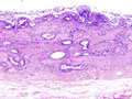
GB carcinoma - low mag. (WC)
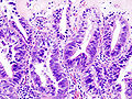
GB carcinoma - high mag. (WC)
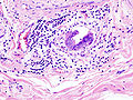
GB carcinoma - LVI. (WC)
_histopathology.jpg)
_lymphatic_invasion_histopathology.jpg)
www:


Moderately differentiated adenocarcinoma of gallbladder in a 59 year old man. A. Tumor bounds cystic duct. B. Cystic duct at left shows high grade dysplasia. At right lie acini and nests of round and sometimes spindled cancerous nuclei. Overall most of the tumor was in tubules/acini, meaning the lesion is moderately differentiated. C. Region with Rokitansky-Aschoff sinuses showing high grade dysplasia and foci of intestinal metaplasia, with some nests likely being invasive cancer. D. Tumorous involvement of cystic duct lymph node. E. Bizarrely shaped cancerous groups. F. invasion of liver by neoplastic acini.
IHC
Features - conventional:[6]
- CK7 +ve (7 of 8 cases).
- CK20 -ve (7 of 8 cases).
- CDX2 -ve (8 of 8 cases).
See also
References
- ↑ Biswas, PK. (Jul 2010). "Carcinoma gallbladder.". Mymensingh Med J 19 (3): 477-81. PMID 20639849.
- ↑ Towfigh S, McFadden DW, Cortina GR, et al (January 2001). "Porcelain gallbladder is not associated with gallbladder carcinoma". Am Surg 67 (1): 7?0. PMID 11206901.
- ↑ Stephen, AE.; Berger, DL. (Jun 2001). "Carcinoma in the porcelain gallbladder: a relationship revisited.". Surgery 129 (6): 699-703. doi:10.1067/msy.2001.113888. PMID 11391368.
- ↑ Kim, HJ.; Park, JH.; Park, DI.; Cho, YK.; Sohn, CI.; Jeon, WK.; Kim, BI.; Choi, SH. (Feb 2012). "Clinical usefulness of endoscopic ultrasonography in the differential diagnosis of gallbladder wall thickening.". Dig Dis Sci 57 (2): 508-15. doi:10.1007/s10620-011-1870-0. PMID 21879282.
- ↑ Tadrous, Paul.J. Diagnostic Criteria Handbook in Histopathology: A Surgical Pathology Vade Mecum (1st ed.). Wiley. pp. 174. ISBN 978-0470519035.
- ↑ 6.0 6.1 Dursun, N.; Escalona, OT.; Roa, JC.; Basturk, O.; Bagci, P.; Cakir, A.; Cheng, J.; Sarmiento, J. et al. (Nov 2012). "Mucinous carcinomas of the gallbladder: clinicopathologic analysis of 15 cases identified in 606 carcinomas.". Arch Pathol Lab Med 136 (11): 1347-58. doi:10.5858/arpa.2011-0447-OA. PMID 23106580.
- ↑ Giang, TH.; Ngoc, TT.; Hassell, LA. (2012). "Carcinoma involving the gallbladder: a retrospective review of 23 cases - pitfalls in diagnosis of gallbladder carcinoma.". Diagn Pathol 7: 10. doi:10.1186/1746-1596-7-10. PMID 22284391.