File:Inked Lumpectomy Specimen (6464023675).jpg

Original file (822 × 627 pixels, file size: 121 KB, MIME type: image/jpeg)
.jpg)
Summary
| Description |
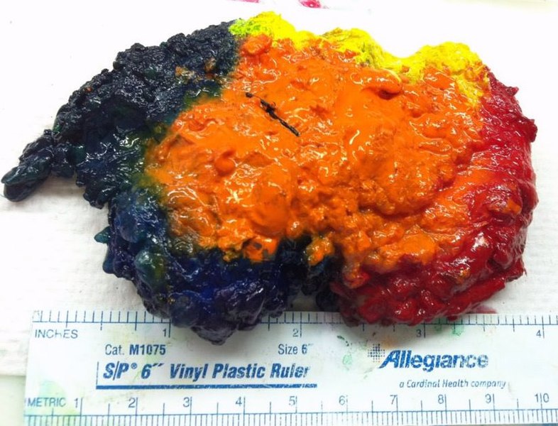
This excisional biopsy was was done for a palpable mass of the left breast. The lesion was considered highly suspicious for malignancy on mammography. Several years previously, the patient had invasive carcinoma of the right breast, with two positive right axillary nodes. At that time, she has an extended simple mastectomy on the right, followed by reconstruction of the right breast and a reduction mammoplasty on the left. She had normal mammograms through 2010, and the abnormality in the left breast suddenly appeared in her 2011 mammogram. Orientation was provided to the pathologist by the surgeon in the operating room. The margins are color-coded: Red-left (lateral) Green-right (medial) Yellow-superior Blue-inferior Orange-anterior Black (not shown on this view)-posterior I took this photo immediately after inking the specimen. Then, I used a squirt bottle to cover the surface with acetone. This dries out the surface and reduces the amount of ink running that occurs when you cut into it. Keep in mind that acetone is volatile and flammable. Other fluids that can be used for the same purpose are Bouin fixative, glacial acetic acid, and white vinegar. Bouin fixative contains picric acid and is therefore a toxic and explosion hazard. On section, the palpable lesion corresponded to a poorly demarcated 1.1-cm hard mass surrounded by normal-appearing tissue. As it turned out, on microscopic examination, the mass was shown to be fat necrosis, perfectly benign. I guess we ended up wasting a lot of time marking the margins on a benign lesion, but unfortunately, it's not possible to tell if a mass is benign until you cut and process it for microscopic exam. Once you cut into it, it's not possible to accurately mark the margins. |
| Date | |
| Source |
|
| Author | Ed Uthman from Houston, TX, USA |
Licensing
- You are free:
- to share – to copy, distribute and transmit the work
- to remix – to adapt the work
- Under the following conditions:
- attribution – You must give appropriate credit, provide a link to the license, and indicate if changes were made. You may do so in any reasonable manner, but not in any way that suggests the licensor endorses you or your use.
| This image, originally posted to Flickr, was reviewed on 11 December 2013 by the administrator or reviewer File Upload Bot (Magnus Manske), who confirmed that it was available on Flickr under the stated license on that date. |
File history
Click on a date/time to view the file as it appeared at that time.
| Date/Time | Thumbnail | Dimensions | User | Comment | |
|---|---|---|---|---|---|
| current | 12:09, 11 December 2013 | | 822 × 627 (121 KB) | File Upload Bot (Magnus Manske) | Transferred from Flickr by User:CFCF |
File usage
The following page uses this file:
.jpg)