Bullous diseases
Bullous diseases are a subset of the large inflammatory skin diseases category. Dermatopathologists help diagnose it.
An introduction to skin pathology is in the dermatopathology article. An introduction to inflammatory skin lesions in the non-malignant skin disease article.
Bullous disease of the lung is dealt with in lung bullae.
Overview
DDx based on type
Subcorneal bullous disorders
DDx with acantholysis:[1]
DDx without acantholysis:DDx:[1]
- Subcorneal pustular dermatosis (Sneddon-Wilkinson disease)
- Pustular psoriasis.
- Pustular drug eruption (acute generalized exanthematous pustulosis).
Suprabasilar bullous disorders
DDx:[1]
- Pemphigus vulgaris.
- Hailey-Hailey disease (benign familial pemphigus).
- Darier disease.
- Grover disease (transient acantholytic dermatosis).
Memory device - PhD + Grover = Pemphigus vulgaris, Hailey-Hailey, Darier, Grover.
Subepidermal bullous disorders
DDx:[1]
- Bullous pemphigoid.
- Cicatricial pemphigoid.
- Porphyria cutanea tarda.
- Epidermolysis bullosa acquista.
- Dermatitis herpetiformis.
- Linear IgA disease.
Others:
- Insect bite.
- Coma blister.
- Bullous systemic lupus erythematosus.
Mnemonic DELPHI:
- Dermatitis herpetiformis.
- Epidermolysis bullosa acquisita.
- Bullous lupus erythematosis.
- Pemphigoid, bullous.
- Herpes gestationis (now called pemphigoid gestationis) - rare autoimmune bullous dermatosis of pregnancy, not related to HSV.[2]
- Linear IgA disease.
Specific diseases
Pemphigus foliaceus
General
- Autoimmune disease.[3]
- Autoantibodies against desmoglein 1 only.
Note:
- Pemphigus vulgaris has autoantibodies against desmoglein 1 and desmoglein 3.[4]
Microscopic
Features:
- Subcorneal separation.
- Acantholysis.
- Separation of keratinocytes.
DDx:
IF
- Desmoglein 1 - abnormal.
Bullous pemphigoid
General
- Less serious than pemphigus vulgaris.
Epidemiology:
- Old people (60-80 year olds).
Clinical:
- Extreme pruritis.
Etiology:
- Antibodies to BPAG2 (a hemidesmosome protein).[5]
Notes:
- Pemphigus vulgaris = subepidermal.
- Pemphigus foliaceus = intraepidermal.
Microscopic
Features:[6]
- Subepidermal blisters.
- +/-Lymphocytes.
- +/-Eosinophils.
- +/-Neutrophils.
Notes:
- Epidermis not affect, i.e. non-acantholytic.
- Linear Ig deposits along basement membrane.
- Early changes may be that of a dermal perivascular lymphoeosinophilic infiltration.
Images:
DDx:
- Bullous lupus.
- Dermal perivascular lymphoeosinophilic infiltration.
Pemphigus vulgaris
- AKA pemphigus.
General
- May lead to blindness.
- Oral lesion is classically: first to show & last to go.
- Oral lesions usually precede the skin lesions.
Classic presentation:
- Mouth lesions.
- Non-pruritic.
Treatment:
- Prednisone then steroid sparing agent.
Epidemiology:
- Associated with thymoma, myasthenia gravis, malignancy & D-penicillamine (used to Tx Wilson's disease).
- Middle age.
Etiology:
Microscopic
Features:[10]
- Suprabasilar blistering.
DDx:
Images:
- PV (dermpedia.org).[11]
- Pemphigus vulgaris - intermed. mag. (WC).
- Pemphigus vulgaris - high mag. (WC).
IF
- Desmoglein 1 - abnormal.
- Desmoglein 3 - abnormal.
Familial benign pemphigus
- AKA Hailey-Hailey disease. Was described by two brothers - that's why it is Hailey-Hailey.[12]
General
- Genetic - autosomal dominant with incomplete penetration.[12]
- Desmosomal defect - due to mutation in the gene ATP2C1.[12]
Clinical:
- Chest.
- Intertriginous regions (only) - where skin rubs together, e.g. skin folds of the breast, axilla.
- Typically presents individual in their 30s and 40s.[12]
Microscopic
Features:
- Suprabasilar blistering.
- Acanthosis (thick epidermis).
Notes:
- Hair folicles spared.
DDx:
Dermatitis herpetiformis
General
- Associated with celiac sprue.
Clinical:
- Pruritis - intense.
Microscopic
Features:[13]
- Subepidermal blistering.
- Clusters of neurophils (microabscesses) - at tips of dermal papillae - key feature.
- Basal cell injury (vacuolization).
Notes:
- Immunofluorescence - IgA deposits at dermal papillae.
Images:
Porphyria cutanea tarda
General
Etiology:
- Genetic, autosomal dominant.
Treatment:
- D/C aggravating substances (see below) - phlebotomy, hydroxychloroquine if phlebotomy contraindicated.
Note:
- Fits into a larger category of porphyria.
Associations
Medications/substances:
Non-infection chronic conditions:
- DM.
Infections:
Gross
- In photoexposed areas subjected to trauma.
Microscopic
Features:[15]
- Subepidermal vesicles.
- Thickening of superficial dermal blood vessels.
Images:
Epidermolysis bullosa acquisita
- Abbreviated EBA
General
- Autoimmune disease.
- Antibodies to collagen type VII.[16]
Microscopic
Features:
- Subepidermal bullae.
Epidermolysis bullosa
General
- A group of inherited, bullous disorders.
- Bullae form due to slight mechanical trauma.
Three major groupings:[17]
- Epidermolysis bullosa simplex (intraepidermal disease).
- Junctional epidermolysis bullosa (separation at DE junction; specifically central portion (lamina lucida)).
- Dystrophic epidermolysis bullosa (separation at DE junction; specifically deep to lamina densa).
Microscopic
Depends on subtype - either intraepidermal or subepidermal.
Grover disease
- AKA transient acantholytic dermatosis.
General
- Cause not known.
- Associated with sun damaged skin, hospitalization and fever.[18]
Clinical:[19]
- Pruritis (itchy).
- Usually self-limited.
- Treatment for symptoms.
Gross
Microscopic
Features:[22]
- Subcorneal bullous disease.
- Acanthosis.
- Dyskeratosis.
DDx:[23]
Images
Grover disease - intermed. mag. (WC)
Grover disease - high mag. (WC)
Grover disease - very high mag. (WC)
Acute generalized exanthematous pustulosis
- Abbreviated AGEP.
- AKA pustular drug eruption.
General
- Drug reaction.
Clinical DDx:
- Erythema multiforme.
- Stevens-Johnson syndrome (SJS) / toxic epidermal necrolysis (TEN).
Microscopic
Features:
- Superficial dermis separates from underlying tissue. (???)
DDx:
Images:
Pemphigoid gestationis
- AKA gestational pemphigoid.
- Previously herpes gestationis.
General
- Autoimmune condition in pregnancy.
- Rare - est. 1/50,000 pregnancies.[26]
Treatment:
- Corticosteroids.[27]
Microscopic
Features:
- Subepidermal bullous disease.[28]
- Eosinophils - abundant.
Images
PG - low mag. (WC)
PG - high mag. (WC)
PG - very high mag. (WC)


IF
- C3 linear pattern at DE junction - diagnostic.[27]
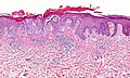
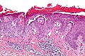
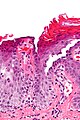
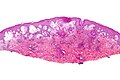
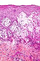
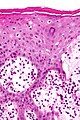
Bullous Arthropod Assault


Bullous arthropod assault. A. Beneath intraepidermal blistering lies superficial and deep inflammation with extension into fat. B. The vesicle shows spongiotic epidermis at base and multiple eosinophils. C. Eosinophils are prominent in dermal and adipose tissue inflammation.
See also
References
- ↑ 1.0 1.1 1.2 1.3 Brinster NK (March 2008). "Dermatopathology for the surgical pathologist: a pattern based approach to the diagnosis of inflammatory skin disorders (part I)". Adv Anat Pathol 15 (2): 76–96. doi:10.1097/PAP.0b013e3181664e8d. PMID 18418089.
- ↑ URL: http://emedicine.medscape.com/article/1063499-overview. Accessed on: 23 September 2011.
- ↑ James, KA.; Culton, DA.; Diaz, LA. (Jul 2011). "Diagnosis and clinical features of pemphigus foliaceus.". Dermatol Clin 29 (3): 405-12, viii. doi:10.1016/j.det.2011.03.012. PMID 21605805.
- ↑ 4.0 4.1 Online 'Mendelian Inheritance in Man' (OMIM) 169615
- ↑ Mitchell, Richard; Kumar, Vinay; Fausto, Nelson; Abbas, Abul K.; Aster, Jon (2011). Pocket Companion to Robbins & Cotran Pathologic Basis of Disease (8th ed.). Elsevier Saunders. pp. 607. ISBN 978-1416054542.
- ↑ Kumar, Vinay; Abbas, Abul K.; Fausto, Nelson; Aster, Jon (2009). Robbins and Cotran pathologic basis of disease (8th ed.). Elsevier Saunders. pp. 1195. ISBN 978-1416031215.
- ↑ URL: http://dermatology.cdlib.org/94/NYU/Feb2002/8.html. Accessed on: 20 March 2011.
- ↑ URL: http://missinglink.ucsf.edu/lm/DermatologyGlossary/bullous_pemphigoid.html. Accessed on: 20 March 2011.
- ↑ Online 'Mendelian Inheritance in Man' (OMIM) 125670
- ↑ Kumar, Vinay; Abbas, Abul K.; Fausto, Nelson; Aster, Jon (2009). Robbins and Cotran pathologic basis of disease (8th ed.). Elsevier Saunders. pp. 1193. ISBN 978-1416031215.
- ↑ URL: http://www.dermpedia.org/baby-dermpedia-for-beginners/pemphigus-vulgaris. Accessed on: 20 March 2011.
- ↑ 12.0 12.1 12.2 12.3 URL: http://emedicine.medscape.com/article/1063224-overview. Accessed on: 9 September 2011.
- ↑ Kumar, Vinay; Abbas, Abul K.; Fausto, Nelson; Aster, Jon (2009). Robbins and Cotran pathologic basis of disease (8th ed.). Elsevier Saunders. pp. 1196. ISBN 978-1416031215.
- ↑ URL: http://dermatology.cdlib.org/94/NYU/Nov2001/9.html. Accessed on: 21 March 2011.
- ↑ Kumar, Vinay; Abbas, Abul K.; Fausto, Nelson; Aster, Jon (2009). Robbins and Cotran pathologic basis of disease (8th ed.). Elsevier Saunders. pp. 1197. ISBN 978-1416031215.
- ↑ URL: http://emedicine.medscape.com/article/1063083-overview. Accessed on: 25 September 2011.
- ↑ URL: http://emedicine.medscape.com/article/1062939-overview. Accessed on: 25 September 2011.
- ↑ Quirk, CJ.; Heenan, PJ. (May 2004). "Grover's disease: 34 years on.". Australas J Dermatol 45 (2): 83-6; quiz 87-8. doi:10.1111/j.1440-0960.2004.054_1.x. PMID 15068451.
- ↑ Hanson, M.; Hsu, S. (Aug 2006). "Pruritic papules on the chest and back. Grover's disease.". Am Fam Physician 74 (4): 641-2. PMID 16939188. http://www.aafp.org/afp/2006/0815/p641.html.
- ↑ Fernández-Figueras, MT.; Puig, L.; Cannata, P.; Cuatrecases, M.; Quer, A.; Ferrándiz, C.; Ariza, A. (Aug 2010). "Grover disease: a reappraisal of histopathological diagnostic criteria in 120 cases.". Am J Dermatopathol 32 (6): 541-9. doi:10.1097/DAD.0b013e3181c80cf9. PMID 20526170.
- ↑ {{Ref APBR|344 Q2
- ↑ S. Sade. 8 September 2011.
- ↑ Davis, MD.; Dinneen, AM.; Landa, N.; Gibson, LE. (Mar 1999). "Grover's disease: clinicopathologic review of 72 cases.". Mayo Clin Proc 74 (3): 229-34. doi:10.4065/74.3.229. PMID 10089990.
- ↑ Intong, LR.; Murrell, DF. (Jul 2011). "Pemphigoid gestationis: pathogenesis and clinical features.". Dermatol Clin 29 (3): 447-52, ix. doi:10.1016/j.det.2011.03.002. PMID 21605810.
- ↑ Tunzi, M.; Gray, GR. (Jan 2007). "Common skin conditions during pregnancy.". Am Fam Physician 75 (2): 211-8. PMID 17263216.
- ↑ Bedocs, PM.; Kumar, V.; Mahon, MJ. (Feb 2009). "Pemphigoid gestationis: a rare case and review.". Arch Gynecol Obstet 279 (2): 235-8. doi:10.1007/s00404-008-0687-3. PMID 18506459.
- ↑ 27.0 27.1 Campbell, SM.; Balazs, K.; Conroy, M. (Jul 2011). "Pemphigoid gestationis: a case report and review of the literature.". Cutis 88 (1): 21-6. PMID 21877502.
- ↑ Kolanko, E.; Bickle, K.; Keehn, C.; Glass, LF. (Mar 2004). "Subepidermal blistering disorders: a clinical and histopathologic review.". Semin Cutan Med Surg 23 (1): 10-8. PMID 15095911.