Difference between revisions of "Venous lake"
Jump to navigation
Jump to search
| (6 intermediate revisions by the same user not shown) | |||
| Line 1: | Line 1: | ||
{{ Infobox diagnosis | |||
| Name = {{PAGENAME}} | |||
| Image = Venous_lake_--_very_low_mag.jpg | |||
| Width = | |||
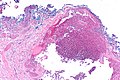
| Caption = Venous lake. [[H&E stain]]. (WC) | |||
| Synonyms = | |||
| Micro = | |||
| Subtypes = | |||
| LMDDx = [[angiokeratoma]], [[cherry hemangioma]] | |||
| Stains = | |||
| IHC = | |||
| EM = | |||
| Molecular = | |||
| IF = | |||
| Gross = classical location: lip | |||
| Grossing = | |||
| Staging = | |||
| Site = | |||
| Assdx = | |||
| Syndromes = | |||
| Clinicalhx = | |||
| Signs = dark blue/purple spot, blanches with pressure | |||
| Symptoms = | |||
| Prevalence = common | |||
| Bloodwork = | |||
| Rads = | |||
| Endoscopy = | |||
| Prognosis = benign | |||
| Other = | |||
| ClinDDx = | |||
| Tx = | |||
}} | |||
'''Venous lake''' is a benign [[skin]] lesion. | '''Venous lake''' is a benign [[skin]] lesion. | ||
| Line 5: | Line 37: | ||
Clinical: | Clinical: | ||
* | *Blanches with pressure.<ref>URL: [http://dermatlas.med.jhmi.edu/derm/IndexDisplay.cfm?ImageID=-969536424 http://dermatlas.med.jhmi.edu/derm/IndexDisplay.cfm?ImageID=-969536424]. Accessed on: 13 August 2012.</ref> | ||
===Gross=== | ===Gross=== | ||
*Purple/blue spot. | *Purple/blue spot. | ||
==Microscopic== | ==Microscopic== | ||
| Line 29: | Line 56: | ||
*[[Cherry hemangioma]].<ref name=Ref_Derm551>{{Ref Derm|551}}</ref> | *[[Cherry hemangioma]].<ref name=Ref_Derm551>{{Ref Derm|551}}</ref> | ||
===Images=== | |||
<gallery> | |||
Image: Venous lake -- very low mag.jpg | VL - very low mag. (WC) | |||
Image: Venous lake -- low mag.jpg | VL - low mag. (WC) | |||
</gallery> | |||
==Sign out== | ==Sign out== | ||
<pre> | |||
Left Lower Lip Lesion, Excision: | |||
- Benign subcutaneous tissue with venous lake. | |||
- NEGATIVE for malignancy. | |||
</pre> | |||
===Block letters=== | |||
<pre> | <pre> | ||
SKIN LESION, RIGHT CHEEK, BIOPSY: | SKIN LESION, RIGHT CHEEK, BIOPSY: | ||
| Line 38: | Line 76: | ||
- NEGATIVE FOR NEVUS. | - NEGATIVE FOR NEVUS. | ||
</pre> | </pre> | ||
===Micro=== | |||
The sections show subcutaneous tissue with dilated vascular spaces. No significant atypia is seen. No overlying skin is present. | |||
==See also== | ==See also== | ||
Latest revision as of 03:06, 23 November 2016
| Venous lake | |
|---|---|
| Diagnosis in short | |
 Venous lake. H&E stain. (WC) | |
| LM DDx | angiokeratoma, cherry hemangioma |
| Gross | classical location: lip |
| Signs | dark blue/purple spot, blanches with pressure |
| Prevalence | common |
| Prognosis | benign |
Venous lake is a benign skin lesion.
General
- Dilated vein.
Clinical:
- Blanches with pressure.[1]
Gross
- Purple/blue spot.
Microscopic
Features:[2]
- Lined by endothelium.
- Blood in lumen.
- +/-Fibrin in lumen.
- +/-Solar elastosis - very common.[3]
DDx:
- Angiokeratoma.
- Ectatic superficial dermal vessels.
- Irregular acanthosis.
- Longer rete ridges.
- Cherry hemangioma.[3]
Images

VL - very low mag. (WC)
VL - low mag. (WC)

Sign out
Left Lower Lip Lesion, Excision:
     - Benign subcutaneous tissue with venous lake.
     - NEGATIVE for malignancy.
Block letters
SKIN LESION, RIGHT CHEEK, BIOPSY:
- VENOUS LAKE.
- SOLAR ELASTOSIS.
- NEGATIVE FOR NEVUS.
Micro
The sections show subcutaneous tissue with dilated vascular spaces. No significant atypia is seen. No overlying skin is present.
See also
References
- ↑ URL: http://dermatlas.med.jhmi.edu/derm/IndexDisplay.cfm?ImageID=-969536424. Accessed on: 13 August 2012.
- ↑ Weedon's Skin Pathology. 3rd Ed. P.895.
- ↑ 3.0 3.1 Busam, Klaus J. (2009). Dermatopathology: A Volume in the Foundations in Diagnostic Pathology Series (1st ed.). Saunders. pp. 551. ISBN 978-0443066542.