Difference between revisions of "Urachal carcinoma"
Jump to navigation
Jump to search
(→IHC) |
|||
| (21 intermediate revisions by the same user not shown) | |||
| Line 3: | Line 3: | ||
| Image = Urachal_carcinoma_-_high_mag.jpg | | Image = Urachal_carcinoma_-_high_mag.jpg | ||
| Width = | | Width = | ||
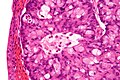
| Caption = Urachal carcinoma. [[H&E stain]]. | | Caption = Urachal carcinoma (right of image) and benign [[urothelium]] (left of image). [[H&E stain]]. | ||
| Micro = atypical cells - usually gland forming, +/-mucinous component, +/-signet rings | | Micro = atypical cells - usually gland forming, +/-mucinous component, +/-signet rings | ||
| Subtypes = enteric, mucinous, signet ring | | Subtypes = enteric, mucinous, signet ring | ||
| LMDDx = [[adenocarcinoma of the urinary bladder]], invasive [[urothelial carcinoma]] with glandular differentiation, [[metastatic]] adenocarcinoma | | LMDDx = [[adenocarcinoma of the urinary bladder]], invasive [[urothelial carcinoma]] with glandular differentiation, [[metastatic]] adenocarcinoma (especially colorectal) | ||
| Stains = | | Stains = | ||
| IHC = CK20 +ve, beta-catenin +ve (non-nuclear), p63 -ve, CK34betaE12 +ve | | IHC = CK20 +ve, beta-catenin +ve (non-nuclear), p63 -ve, CK34betaE12 +ve | ||
| Line 13: | Line 13: | ||
| IF = | | IF = | ||
| Gross = | | Gross = | ||
| Grossing = | | Grossing = [[partial cystectomy]] | ||
| Site = urachus, [[urinary bladder]] - specifically the dome | | Site = urachus, [[urinary bladder]] - specifically the dome | ||
| Assdx = | | Assdx = | ||
| Syndromes = | | Syndromes = | ||
| Clinicalhx = | | Clinicalhx = no prior urothelial carcinoma | ||
| Signs = +/-hematuria | | Signs = +/-hematuria | ||
| Symptoms = | | Symptoms = | ||
| Prevalence = very rare | | Prevalence = very rare | ||
| Bloodwork = | | Bloodwork = | ||
| Rads = | | Rads = lesion of bladder dome/anterior bladder, lesion centered in wall of bladder (rather than mucosa) | ||
| Endoscopy = | | Endoscopy = | ||
| Prognosis = usually poor | | Prognosis = usually poor | ||
| Line 29: | Line 29: | ||
}} | }} | ||
'''Urachal carcinoma''' is an uncommon [[cancer|malignant tumour]] that arises from the urachus. Most urachal carcinomas are ''[[adenocarcinoma]]s''. | '''Urachal carcinoma''' is an uncommon [[cancer|malignant tumour]] that arises from the urachus. Most urachal carcinomas are ''[[adenocarcinoma]]s''. | ||
The diagnosis is a clinicopathologic diagnosis - one needs imaging to make it.<ref name=Ref_Amin2-143>{{Ref Amin|2-143}}</ref> | |||
==General== | ==General== | ||
*Very rare<ref name=pmid16826585>{{Cite journal | last1 = Ashley | first1 = RA. | last2 = Inman | first2 = BA. | last3 = Sebo | first3 = TJ. | last4 = Leibovich | first4 = BC. | last5 = Blute | first5 = ML. | last6 = Kwon | first6 = ED. | last7 = Zincke | first7 = H. | title = Urachal carcinoma: clinicopathologic features and long-term outcomes of an aggressive malignancy. | journal = Cancer | volume = 107 | issue = 4 | pages = 712-20 | month = Aug | year = 2006 | doi = 10.1002/cncr.22060 | PMID = 16826585 }}</ref>~ 0.2% of bladder cancers.<ref name=pmid22901574>{{Cite journal | last1 = Bruins | first1 = HM. | last2 = Visser | first2 = O. | last3 = Ploeg | first3 = M. | last4 = Hulsbergen-van de Kaa | first4 = CA. | last5 = Kiemeney | first5 = LA. | last6 = Witjes | first6 = JA. | title = The clinical epidemiology of urachal carcinoma: results of a large, population based study. | journal = J Urol | volume = 188 | issue = 4 | pages = 1102-7 | month = Oct | year = 2012 | doi = 10.1016/j.juro.2012.06.020 | PMID = 22901574 }}</ref> | *Very rare<ref name=pmid16826585>{{Cite journal | last1 = Ashley | first1 = RA. | last2 = Inman | first2 = BA. | last3 = Sebo | first3 = TJ. | last4 = Leibovich | first4 = BC. | last5 = Blute | first5 = ML. | last6 = Kwon | first6 = ED. | last7 = Zincke | first7 = H. | title = Urachal carcinoma: clinicopathologic features and long-term outcomes of an aggressive malignancy. | journal = Cancer | volume = 107 | issue = 4 | pages = 712-20 | month = Aug | year = 2006 | doi = 10.1002/cncr.22060 | PMID = 16826585 }}</ref>~ 0.2% of bladder cancers.<ref name=pmid22901574>{{Cite journal | last1 = Bruins | first1 = HM. | last2 = Visser | first2 = O. | last3 = Ploeg | first3 = M. | last4 = Hulsbergen-van de Kaa | first4 = CA. | last5 = Kiemeney | first5 = LA. | last6 = Witjes | first6 = JA. | title = The clinical epidemiology of urachal carcinoma: results of a large, population based study. | journal = J Urol | volume = 188 | issue = 4 | pages = 1102-7 | month = Oct | year = 2012 | doi = 10.1016/j.juro.2012.06.020 | PMID = 22901574 }}</ref> | ||
* | *Often younger <55 years-old. | ||
*Poor prognosis<ref name=pmid24136277/> - especially for high [[Sheldon stage]].<ref name=pmid19252435>{{Cite journal | last1 = Gopalan | first1 = A. | last2 = Sharp | first2 = DS. | last3 = Fine | first3 = SW. | last4 = Tickoo | first4 = SK. | last5 = Herr | first5 = HW. | last6 = Reuter | first6 = VE. | last7 = Olgac | first7 = S. | title = Urachal carcinoma: a clinicopathologic analysis of 24 cases with outcome correlation. | journal = Am J Surg Pathol | volume = 33 | issue = 5 | pages = 659-68 | month = May | year = 2009 | doi = 10.1097/PAS.0b013e31819aa4ae | PMID = 19252435 }}</ref> | |||
Clinical: | |||
*Hematuria - classic presentation.<ref name=pmid24136277>{{Cite journal | last1 = Shao | first1 = GJ. | last2 = Cai | first2 = L. | last3 = Li | first3 = XS. | last4 = Song | first4 = G. | last5 = Li | first5 = XY. | last6 = He | first6 = ZS. | last7 = Zhou | first7 = LQ. | title = [Urachal carcinoma: experience of a clinical center within 30 years]. | journal = Beijing Da Xue Xue Bao | volume = 45 | issue = 5 | pages = 774-8 | month = Oct | year = 2013 | doi = | PMID = 24136277 }}</ref> | |||
Treatment: | Treatment: | ||
*Partial cystectomy +/- umbilectomy. | *[[Partial cystectomy]] +/- umbilectomy. | ||
==Gross== | ==Gross== | ||
| Line 41: | Line 47: | ||
==Microscopic== | ==Microscopic== | ||
Features: | Features of epithelial malignancy: | ||
*Atypical cells. | *Atypical (epithelial) cells. | ||
**May be signet ring cells. | **May be [[signet ring cells]]. | ||
*Usually gland forming, i.e. adenocarcinoma. | *Usually gland forming, i.e. adenocarcinoma. | ||
*+/-Mucinous component. | *+/-Mucinous component. | ||
* | Features making it ''urachal carcinoma'' (modified Sheldon criteria) - all required:<ref name=pmid22301493>{{Cite journal | last1 = Paner | first1 = GP. | last2 = Barkan | first2 = GA. | last3 = Mehta | first3 = V. | last4 = Sirintrapun | first4 = SJ. | last5 = Tsuzuki | first5 = T. | last6 = Sebo | first6 = TJ. | last7 = Jimenez | first7 = RE. | title = Urachal carcinomas of the nonglandular type: salient features and considerations in pathologic diagnosis. | journal = Am J Surg Pathol | volume = 36 | issue = 3 | pages = 432-42 | month = Mar | year = 2012 | doi = 10.1097/PAS.0b013e31823fe49c | PMID = 22301493 }}</ref> | ||
#Located in bladder dome or anterior wall. † | |||
#Centered on bladder wall (as opposed to the mucosa). | |||
#No widespread cystitis cystica/glandularis outside of the bladder dome and anterior wall. | |||
#No known urothelial carcinoma elsewhere. | |||
Notes: | |||
*Gopalan ''et al.''<ref name=pmid19252435/> (based on Sheldon) suggests the following additional/more restrictive criteria: | |||
**Only the dome is allowed. † | |||
**Lesion should be sharply demarcated from the [[urothelium]] of the urinary bladder. | |||
**Urachal remnants should be present in the tumour. | |||
DDx:<ref name=Ref_Amin2-143>{{Ref Amin|2-143}}</ref> | DDx:<ref name=Ref_Amin2-143>{{Ref Amin|2-143}}</ref> | ||
*[[Adenocarcinoma of the urinary bladder]]. | *[[Adenocarcinoma of the urinary bladder]]. | ||
*Invasive [[urothelial carcinoma]] with glandular differentiation. | *Invasive [[urothelial carcinoma]] with glandular differentiation. | ||
*Metastatic adenocarcinoma/adenocarcinoma extending from another structure, e.g. [[colorectal adenocarcinoma]]. | *[[Metastatic]] adenocarcinoma/[[adenocarcinoma]] extending from another structure, e.g. [[colorectal adenocarcinoma]]. | ||
*Villous adenoma of the urachus<ref>{{Cite journal | last1 = Joniau | first1 = S. | last2 = Lerut | first2 = E. | last3 = Van Poppel | first3 = H. | title = A Giant Mucinous Adenocarcinoma Arising within a Villous Adenoma of the Urachus: Case Report and Review of the Literature. | journal = Case Rep Med | volume = 2009 | issue = | pages = 818646 | month = | year = 2009 | doi = 10.1155/2009/818646 | PMID = 20182635 }}</ref> - non-invasive. | |||
===Patterns=== | ===Patterns=== | ||
*Enteric - looks like colonic adenocarcinoma. | *Enteric - looks like colonic adenocarcinoma. | ||
*Mucinous. | *Mucinous. | ||
*Signet ring. | *[[signet ring cell carcinoma|Signet ring]]. | ||
Note: | Note: | ||
| Line 62: | Line 79: | ||
===Images=== | ===Images=== | ||
<gallery> | <gallery> | ||
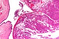
Image: | Image: Urachal carcinoma - very low mag.jpg | UC - very low mag. (WC/Nephron) | ||
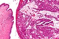
Image: | Image: Urachal carcinoma - low mag.jpg | UC - low mag. (WC/Nephron) | ||
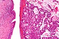
Image: | Image: Urachal carcinoma - intermed mag.jpg | UC - intermed mag. (WC/Nephron) | ||
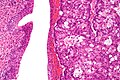
Image: Urachal carcinoma - high mag.jpg | UC - high mag. (WC/Nephron) | |||
Image: Urachal carcinoma - very high mag.jpg | UC - very high mag. (WC/Nephron) | |||
</gallery> | </gallery> | ||
==IHC== | ==IHC== | ||
Features:<ref name=pmid19252435>{{Cite journal | last1 = Gopalan | first1 = A. | last2 = Sharp | first2 = DS. | last3 = Fine | first3 = SW. | last4 = Tickoo | first4 = SK. | last5 = Herr | first5 = HW. | last6 = Reuter | first6 = VE. | last7 = Olgac | first7 = S. | title = Urachal carcinoma: a clinicopathologic analysis of 24 cases with outcome correlation. | journal = Am J Surg Pathol | volume = 33 | issue = 5 | pages = 659-68 | month = May | year = 2009 | doi = 10.1097/PAS.0b013e31819aa4ae | PMID = 19252435 }}</ref> | Features:<ref name=pmid19252435>{{Cite journal | last1 = Gopalan | first1 = A. | last2 = Sharp | first2 = DS. | last3 = Fine | first3 = SW. | last4 = Tickoo | first4 = SK. | last5 = Herr | first5 = HW. | last6 = Reuter | first6 = VE. | last7 = Olgac | first7 = S. | title = Urachal carcinoma: a clinicopathologic analysis of 24 cases with outcome correlation. | journal = Am J Surg Pathol | volume = 33 | issue = 5 | pages = 659-68 | month = May | year = 2009 | doi = 10.1097/PAS.0b013e31819aa4ae | PMID = 19252435 }}</ref> | ||
*CK20 +ve. | *[[CK20]] +ve. | ||
*CK7 +ve/-ve. | *[[CK7]] +ve/-ve. | ||
*CK34betaE12 +ve/-ve. | *[[CK34betaE12]] +ve/-ve. | ||
*Beta-catenin -- usu cytoplasmic/membranous +ve. | *Beta-catenin -- usu cytoplasmic/membranous +ve. | ||
Others:<ref name=pmid21572312>{{Cite journal | last1 = Paner | first1 = GP. | last2 = McKenney | first2 = JK. | last3 = Barkan | first3 = GA. | last4 = Yao | first4 = JL. | last5 = Frankel | first5 = WL. | last6 = Sebo | first6 = TJ. | last7 = Shen | first7 = SS. | last8 = Jimenez | first8 = RE. | title = Immunohistochemical analysis in a morphologic spectrum of urachal epithelial neoplasms: diagnostic implications and pitfalls. | journal = Am J Surg Pathol | volume = 35 | issue = 6 | pages = 787-98 | month = Jun | year = 2011 | doi = 10.1097/PAS.0b013e3182189c11 | PMID = 21572312 }}</ref> | Others:<ref name=pmid21572312>{{Cite journal | last1 = Paner | first1 = GP. | last2 = McKenney | first2 = JK. | last3 = Barkan | first3 = GA. | last4 = Yao | first4 = JL. | last5 = Frankel | first5 = WL. | last6 = Sebo | first6 = TJ. | last7 = Shen | first7 = SS. | last8 = Jimenez | first8 = RE. | title = Immunohistochemical analysis in a morphologic spectrum of urachal epithelial neoplasms: diagnostic implications and pitfalls. | journal = Am J Surg Pathol | volume = 35 | issue = 6 | pages = 787-98 | month = Jun | year = 2011 | doi = 10.1097/PAS.0b013e3182189c11 | PMID = 21572312 }}</ref> | ||
*p63 -ve (+ve in only 3%). | *[[p63]] -ve (+ve in only 3%). | ||
UC versus [[CRC]] -- not absolute but useful: | UC versus [[CRC]] -- not absolute but useful: | ||
| Line 82: | Line 101: | ||
==Sign out== | ==Sign out== | ||
<pre> | |||
Mass, Urachus, Resection: | |||
- ADENOCARCINOMA, consistent with urachal primary, see comment. | |||
-- Margins clear. | |||
Comment: | |||
The sections show malignant cells within the perivesicular soft tissue arranged in nests and glands with necrosis. The lesion center is within the bladder wall, rather than at the mucosa. The sampled bladder mucosa is within normal limits. | |||
The tumour stains as follows: | |||
POSITIVE: CDX2 (focal, moderate nuclear), CK20 (focal, moderate membranous). | |||
NEGATIVE: CK7, p63, CK5/6, GATA3, beta-catenin (membranous only, nuclei negative). | |||
The histomorphology, immunoprofile and imaging are compatible with a primary urachal adenocarcinoma. | |||
The morphology and immunoprofile of urachal adenocarcinoma overlaps with primary adenocarcinoma of the bladder and somewhat with colorectal adenocarcinoma. Primary adenocarcinoma of the bladder is unlikely as the sampled urothelium is benign, the lesion is centered within the wall and no history of bladder tumours was found. Metastatic colorectal cancer seems unlikely from the clinical information available. | |||
The CAP bladder synoptic is not applicable to urachal carcinoma. | |||
In the context of the "Sheldon Staging System" for urachal carcinoma, this case would be pT3a. | |||
</pre> | |||
===Staging=== | |||
*The CAP urinary bladder synoptic, as per v4.0.2.0, should ''not'' be used. | |||
*May be [[cancer staging|staged]] with the ''Sheldon system''.<ref name=pmid22901574>{{Cite journal | last1 = Bruins | first1 = HM. | last2 = Visser | first2 = O. | last3 = Ploeg | first3 = M. | last4 = Hulsbergen-van de Kaa | first4 = CA. | last5 = Kiemeney | first5 = LA. | last6 = Witjes | first6 = JA. | title = The clinical epidemiology of urachal carcinoma: results of a large, population based study. | journal = J Urol | volume = 188 | issue = 4 | pages = 1102-7 | month = Oct | year = 2012 | doi = 10.1016/j.juro.2012.06.020 | PMID = 22901574 }}</ref> | *May be [[cancer staging|staged]] with the ''Sheldon system''.<ref name=pmid22901574>{{Cite journal | last1 = Bruins | first1 = HM. | last2 = Visser | first2 = O. | last3 = Ploeg | first3 = M. | last4 = Hulsbergen-van de Kaa | first4 = CA. | last5 = Kiemeney | first5 = LA. | last6 = Witjes | first6 = JA. | title = The clinical epidemiology of urachal carcinoma: results of a large, population based study. | journal = J Urol | volume = 188 | issue = 4 | pages = 1102-7 | month = Oct | year = 2012 | doi = 10.1016/j.juro.2012.06.020 | PMID = 22901574 }}</ref> | ||
====Sheldon staging system==== | |||
Staging as per Sheldon ''et al.'':<ref name=pmid19252435>{{Cite journal | last1 = Gopalan | first1 = A. | last2 = Sharp | first2 = DS. | last3 = Fine | first3 = SW. | last4 = Tickoo | first4 = SK. | last5 = Herr | first5 = HW. | last6 = Reuter | first6 = VE. | last7 = Olgac | first7 = S. | title = Urachal carcinoma: a clinicopathologic analysis of 24 cases with outcome correlation. | journal = Am J Surg Pathol | volume = 33 | issue = 5 | pages = 659-68 | month = May | year = 2009 | doi = 10.1097/PAS.0b013e31819aa4ae | PMID = 19252435 }}</ref><ref name=pmid6361280>{{Cite journal | last1 = Sheldon | first1 = CA. | last2 = Clayman | first2 = RV. | last3 = Gonzalez | first3 = R. | last4 = Williams | first4 = RD. | last5 = Fraley | first5 = EE. | title = Malignant urachal lesions. | journal = J Urol | volume = 131 | issue = 1 | pages = 1-8 | month = Jan | year = 1984 | doi = | PMID = 6361280 }}</ref> | |||
*pT1 - confined to urachal mucosa. | |||
*pT2 - confined to the urachus. | |||
*pT3a - extension into the bladder wall | |||
*pT3b - extension into the abdominal wall. | |||
*pT3c - structures other than the bladder. | |||
*pT4a - metastasis to regional [[lymph node]]s. | |||
*pT4b - metastasis to distant sites. | |||
==See also== | ==See also== | ||
Latest revision as of 14:54, 4 August 2022
| Urachal carcinoma | |
|---|---|
| Diagnosis in short | |
 Urachal carcinoma (right of image) and benign urothelium (left of image). H&E stain. | |
|
| |
| LM | atypical cells - usually gland forming, +/-mucinous component, +/-signet rings |
| Subtypes | enteric, mucinous, signet ring |
| LM DDx | adenocarcinoma of the urinary bladder, invasive urothelial carcinoma with glandular differentiation, metastatic adenocarcinoma (especially colorectal) |
| IHC | CK20 +ve, beta-catenin +ve (non-nuclear), p63 -ve, CK34betaE12 +ve |
| Grossing notes | partial cystectomy |
| Site | urachus, urinary bladder - specifically the dome |
|
| |
| Clinical history | no prior urothelial carcinoma |
| Signs | +/-hematuria |
| Prevalence | very rare |
| Radiology | lesion of bladder dome/anterior bladder, lesion centered in wall of bladder (rather than mucosa) |
| Prognosis | usually poor |
| Clin. DDx | other bladder tumours - esp. urothelial carcinoma |
Urachal carcinoma is an uncommon malignant tumour that arises from the urachus. Most urachal carcinomas are adenocarcinomas.
The diagnosis is a clinicopathologic diagnosis - one needs imaging to make it.[1]
General
- Very rare[2]~ 0.2% of bladder cancers.[3]
- Often younger <55 years-old.
- Poor prognosis[4] - especially for high Sheldon stage.[5]
Clinical:
- Hematuria - classic presentation.[4]
Treatment:
- Partial cystectomy +/- umbilectomy.
Gross
- Lesion must be in urachus or dome of urinary bladder.
Microscopic
Features of epithelial malignancy:
- Atypical (epithelial) cells.
- May be signet ring cells.
- Usually gland forming, i.e. adenocarcinoma.
- +/-Mucinous component.
Features making it urachal carcinoma (modified Sheldon criteria) - all required:[6]
- Located in bladder dome or anterior wall. †
- Centered on bladder wall (as opposed to the mucosa).
- No widespread cystitis cystica/glandularis outside of the bladder dome and anterior wall.
- No known urothelial carcinoma elsewhere.
Notes:
- Gopalan et al.[5] (based on Sheldon) suggests the following additional/more restrictive criteria:
- Only the dome is allowed. †
- Lesion should be sharply demarcated from the urothelium of the urinary bladder.
- Urachal remnants should be present in the tumour.
DDx:[1]
- Adenocarcinoma of the urinary bladder.
- Invasive urothelial carcinoma with glandular differentiation.
- Metastatic adenocarcinoma/adenocarcinoma extending from another structure, e.g. colorectal adenocarcinoma.
- Villous adenoma of the urachus[7] - non-invasive.
Patterns
- Enteric - looks like colonic adenocarcinoma.
- Mucinous.
- Signet ring.
Note:
- Urachal carcinoma may be nonglandular.[6]
Images
UC - very low mag. (WC/Nephron)
UC - low mag. (WC/Nephron)
UC - intermed mag. (WC/Nephron)
UC - high mag. (WC/Nephron)
UC - very high mag. (WC/Nephron)


IHC
Features:[5]
- CK20 +ve.
- CK7 +ve/-ve.
- CK34betaE12 +ve/-ve.
- Beta-catenin -- usu cytoplasmic/membranous +ve.
Others:[8]
- p63 -ve (+ve in only 3%).
UC versus CRC -- not absolute but useful:
- CK34betaE12 +ve in UC (-ve in CRC).
- Beta-catenin -ve nuclei in UC (+ve nuclei in CRC).
Sign out
Mass, Urachus, Resection:
     - ADENOCARCINOMA, consistent with urachal primary, see comment.
     -- Margins clear.

Comment:
The sections show malignant cells within the perivesicular soft tissue arranged in nests and glands with necrosis. The lesion center is within the bladder wall, rather than at the mucosa. The sampled bladder mucosa is within normal limits.

The tumour stains as follows:
POSITIVE: CDX2 (focal, moderate nuclear), CK20 (focal, moderate membranous).
NEGATIVE: CK7, p63, CK5/6, GATA3, beta-catenin (membranous only, nuclei negative).

The histomorphology, immunoprofile and imaging are compatible with a primary urachal adenocarcinoma.

The morphology and immunoprofile of urachal adenocarcinoma overlaps with primary adenocarcinoma of the bladder and somewhat with colorectal adenocarcinoma. Primary adenocarcinoma of the bladder is unlikely as the sampled urothelium is benign, the lesion is centered within the wall and no history of bladder tumours was found.  Metastatic colorectal cancer seems unlikely from the clinical information available.

The CAP bladder synoptic is not applicable to urachal carcinoma.

In the context of the "Sheldon Staging System" for urachal carcinoma, this case would be pT3a.
Staging
- The CAP urinary bladder synoptic, as per v4.0.2.0, should not be used.
- May be staged with the Sheldon system.[3]
Sheldon staging system
Staging as per Sheldon et al.:[5][9]
- pT1 - confined to urachal mucosa.
- pT2 - confined to the urachus.
- pT3a - extension into the bladder wall
- pT3b - extension into the abdominal wall.
- pT3c - structures other than the bladder.
- pT4a - metastasis to regional lymph nodes.
- pT4b - metastasis to distant sites.
See also
References
- ↑ 1.0 1.1 Amin, Mahul B. (2010). Diagnostic Pathology: Genitourinary (1st ed.). Amirsys. pp. 2-143. ISBN 978-1931884280.
- ↑ Ashley, RA.; Inman, BA.; Sebo, TJ.; Leibovich, BC.; Blute, ML.; Kwon, ED.; Zincke, H. (Aug 2006). "Urachal carcinoma: clinicopathologic features and long-term outcomes of an aggressive malignancy.". Cancer 107 (4): 712-20. doi:10.1002/cncr.22060. PMID 16826585.
- ↑ 3.0 3.1 Bruins, HM.; Visser, O.; Ploeg, M.; Hulsbergen-van de Kaa, CA.; Kiemeney, LA.; Witjes, JA. (Oct 2012). "The clinical epidemiology of urachal carcinoma: results of a large, population based study.". J Urol 188 (4): 1102-7. doi:10.1016/j.juro.2012.06.020. PMID 22901574.
- ↑ 4.0 4.1 Shao, GJ.; Cai, L.; Li, XS.; Song, G.; Li, XY.; He, ZS.; Zhou, LQ. (Oct 2013). "[Urachal carcinoma: experience of a clinical center within 30 years].". Beijing Da Xue Xue Bao 45 (5): 774-8. PMID 24136277.
- ↑ 5.0 5.1 5.2 5.3 Gopalan, A.; Sharp, DS.; Fine, SW.; Tickoo, SK.; Herr, HW.; Reuter, VE.; Olgac, S. (May 2009). "Urachal carcinoma: a clinicopathologic analysis of 24 cases with outcome correlation.". Am J Surg Pathol 33 (5): 659-68. doi:10.1097/PAS.0b013e31819aa4ae. PMID 19252435.
- ↑ 6.0 6.1 Paner, GP.; Barkan, GA.; Mehta, V.; Sirintrapun, SJ.; Tsuzuki, T.; Sebo, TJ.; Jimenez, RE. (Mar 2012). "Urachal carcinomas of the nonglandular type: salient features and considerations in pathologic diagnosis.". Am J Surg Pathol 36 (3): 432-42. doi:10.1097/PAS.0b013e31823fe49c. PMID 22301493.
- ↑ Joniau, S.; Lerut, E.; Van Poppel, H. (2009). "A Giant Mucinous Adenocarcinoma Arising within a Villous Adenoma of the Urachus: Case Report and Review of the Literature.". Case Rep Med 2009: 818646. doi:10.1155/2009/818646. PMID 20182635.
- ↑ Paner, GP.; McKenney, JK.; Barkan, GA.; Yao, JL.; Frankel, WL.; Sebo, TJ.; Shen, SS.; Jimenez, RE. (Jun 2011). "Immunohistochemical analysis in a morphologic spectrum of urachal epithelial neoplasms: diagnostic implications and pitfalls.". Am J Surg Pathol 35 (6): 787-98. doi:10.1097/PAS.0b013e3182189c11. PMID 21572312.
- ↑ Sheldon, CA.; Clayman, RV.; Gonzalez, R.; Williams, RD.; Fraley, EE. (Jan 1984). "Malignant urachal lesions.". J Urol 131 (1): 1-8. PMID 6361280.