Testicular atrophy
| Testicular atrophy | |
|---|---|
| Diagnosis in short | |
 Atrophic changes of the testis (bottom). H&E stain. | |
|
| |
| LM | thickening of seminiferous tubule basement membrane, decreased sperm/no sperm present, +/-intertubular fibrosis |
| Site | testis |
|
| |
| Clinical history | undescended testis (cryptorchidism) |
| Prevalence | not common |
| Prognosis | benign |
| Clin. DDx | dependent on history, ?testicular mass |
| Treatment | none required |
Testicular atrophy is relatively common change seen in undescended testes. It is also known as atrophic testis and atrophy of the testis.
Cryptorchidism redirects here.
General
- Microscopic appearance identical to cryptorchidism (undescended testis).[1]
Gross
- Decreased size.
Microscopic
Features:[1]
- Thickening of seminiferous tubule basement membrane.
- Intertubular fibrosis.
- Decreased sperm/no sperm present.
Note:
- End-stage testicle - only has Sertoli cell within the seminiferous tubules.
Other considerations (DDx):
Images
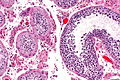
Atrophic changes (left) - intermed. mag.
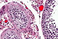
Atrophic changes (left) - high mag.
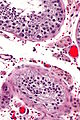
Atrophic changes (bottom) - high mag.
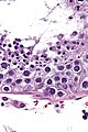
Testis - very high mag.


www:
Sign out
TESTICLE, RIGHT, ORCHIECTOMY:
- ATROPHIC TESTICLE.
- NEGATIVE FOR INTRATUBULAR GERM CELL NEOPLASIA.
- NEGATIVE FOR MALIGNANCY.
Micro
The sections show seminiferous tubules surrounded by thick hyaline sleeves. In a large number of sections only Sertoli cells are found in the tubules.
In some sections poorly defined paucicellular tubular structures reminiscent of seminiferous tubules composed of hyaline material are present; these probably represent obsolete seminiferous tubules. Focally, fibrosis is seen without definite tumour outlines. There is no significant inflammation. The rete testis is identified.
Rare seminiferous tubules have spermatid within. The germ cells seen do not have appreciable nuclear atypia.
Numerous small Leydig cell clusters are seen in some sections.
See also
- Testis.
- Pick's adenoma (Sertoli cell nodule).
References
- ↑ 1.0 1.1 Mitchell, Richard; Kumar, Vinay; Fausto, Nelson; Abbas, Abul K.; Aster, Jon (2011). Pocket Companion to Robbins & Cotran Pathologic Basis of Disease (8th ed.). Elsevier Saunders. pp. 506-7. ISBN 978-1416054542.