Difference between revisions of "Terminal bar"
Jump to navigation
Jump to search

| Line 14: | Line 14: | ||
==Related images== | ==Related images== | ||
<gallery> | <gallery> | ||
Image: Bronchial cells - bronchial wash -- very high mag.jpg | | Image: Bronchial cells - bronchial wash -- very high mag.jpg | BCs - high mag. | ||
Image: Bronchial cells - bronchial wash - 2 -- very high mag.jpg | | Image: Bronchial cells - bronchial wash - 2 -- very high mag.jpg | BCs - high mag. | ||
Image: Bronchial cells - bronchial wash - 2 cropped -- very high mag.jpg | | Image: Bronchial cells - bronchial wash - 2 cropped -- very high mag.jpg | BCs - high mag. | ||
</gallery> | </gallery> | ||
Latest revision as of 05:19, 26 October 2015

Terminal bar in bronchial cells. Pap stain. (WC)
The terminal bar is a cytologic finding in ciliated cells that suggests benignancy.
General
- Terminal bar present = strong indicator that the cell is benign.[1]
Cytology
Features:
- Dense band-like region in the luminal aspect of the cell.
- Dark blue colour on Pap stain.
- Typically seen together with cilia.
Related images
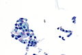
BCs - high mag.
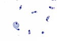
BCs - high mag.
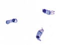
BCs - high mag.


See also
References
- ↑ Van Le, L.; Novotny, D.; Dotters, DJ. (Nov 1991). "Distinguishing tubal metaplasia from endocervical dysplasia on cervical Papanicolaou smears.". Obstet Gynecol 78 (5 Pt 2): 974-6. PMID 1923244.
- ↑ Sciallis, AP.; Aubry, MC.; Bell, DA. (Sep 2009). "Ciliated adenocarcinoma of the ovary with evidence of serous differentiation: report of a case.". Int J Gynecol Pathol 28 (5): 447-52. doi:10.1097/PGP.0b013e3181a0717f. PMID 19696614.