Subependymal giant cell astrocytoma
Jump to navigation
Jump to search
The printable version is no longer supported and may have rendering errors. Please update your browser bookmarks and please use the default browser print function instead.
| Subependymal giant cell astrocytoma | |
|---|---|
| Diagnosis in short | |
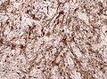
 Subependymal giant cell astrocytoma H&E stain. | |
|
| |
| Synonyms | SEGA |
| LM DDx | ganglioglioma, pleomorphic xanthoastrocytoma, glioblastoma |
| IHC | GFAP +ve |
| Site | brain - usu. wall of ventricles |
|
| |
| Prevalence | rare - esp. in young adults |
| Prognosis | good (WHO Grade I) |
Subependymal giant cell astrocytoma, abbreviated SEGA, is a low-grade astrocytoma associated with tuberous sclerosis complex.
General
- Associated with tuberous sclerosis complex (TSC).[1]
- 6-14% of all TSC patients will develop a SEGA.
- Sporadic examples of SEGA may represent undetected TSC patients (i.e., low-level somatic mosaicism)[2].
- Associated with epilepsy.
- WHO Grade I.
Gross/radiology
- Well-demarcated.
- Often projecting into a ventricle.
- May be calcified
- Circumscribed tumour.
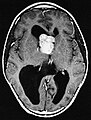
SEGA in Tuberous sclerosis. (WC/AFIP)
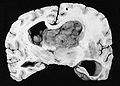
SEGA. (WC/AFIP)


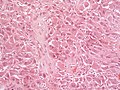
Microscopic
- Giant cells with nuclear atypia ("bizarre cells", "ganglioid cells").
- Glassy eosinophilic cytoplasm.
- Elongated cells in a fibrillary background.
- Abundant mast cells.[5]
- Lymphocytic infiltrates.
- Endothelial proliferations and/or necrosis are not a sign of malignancy.
Images
SEGA. (WC/Sbrandner)
SEGA - NF stain. (WC/Sbrandner)
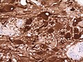
SEGA - GFAP stain. (WC/jensflorian)
SEGA -GFAP stain. (WC/Sbrandner)


www:
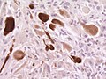
IHC
- GFAP +ve. (50%)
- Vimentin +ve. (100%)
- S100 +ve. (100%)
- Neurofilament +/-ve (ganglionic component).
- Synaptophysin +/-ve (ganglionic component)..
- TTF-1 (7 out of 7).[7]
- Olig2-ve.[8]
- MIB-1 usu. low (1-5%).
See also
References
- ↑ Grajkowska, W.; Kotulska, K.; Jurkiewicz, E.; Roszkowski, M.; Daszkiewicz, P.; Jóźwiak, S.; Matyja, E. (2011). "Subependymal giant cell astrocytomas with atypical histological features mimicking malignant gliomas.". Folia Neuropathol 49 (1): 39-46. PMID 21455842.
- ↑ Overwater, IE.; Swenker, R.; van der Ende, EL.; Hanemaayer, KB.; Hoogeveen-Westerveld, M.; van Eeghen, AM.; Lequin, MH.; van den Ouweland, AM. et al. (12 2016). "Genotype and brain pathology phenotype in children with tuberous sclerosis complex.". Eur J Hum Genet 24 (12): 1688-1695. doi:10.1038/ejhg.2016.85. PMID 27406250.
- ↑ 3.0 3.1 URL: http://path.upmc.edu/cases/case179.html. Accessed on: 29 July 2011.
- ↑ 4.0 4.1 Taraszewska, A.; Kroh, H.; Majchrowski, A. (1997). "Subependymal giant cell astrocytoma: clinical, histologic and immunohistochemical characteristic of 3 cases.". Folia Neuropathol 35 (3): 181-6. PMID 9595853.
- ↑ 5.0 5.1 URL: http://path.upmc.edu/cases/case179/micro.html. Accessed on: 8 January 2012.
- ↑ Hirose, T.; Scheithauer, BW.; Lopes, MB.; Gerber, HA.; Altermatt, HJ.; Hukee, MJ.; VandenBerg, SR.; Charlesworth, JC. (1995). "Tuber and subependymal giant cell astrocytoma associated with tuberous sclerosis: an immunohistochemical, ultrastructural, and immunoelectron and microscopic study.". Acta Neuropathol 90 (4): 387-99. PMID 8546029.
- ↑ Hewer, E.; Vajtai, I.. "Consistent nuclear expression of thyroid transcription factor 1 in subependymal giant cell astrocytomas suggests lineage-restricted histogenesis.". Clin Neuropathol 34 (3): 128-31. doi:10.5414/NP300818. PMID 25669749.
- ↑ Overwater, IE.; Swenker, R.; van der Ende, EL.; Hanemaayer, KB.; Hoogeveen-Westerveld, M.; van Eeghen, AM.; Lequin, MH.; van den Ouweland, AM. et al. (12 2016). "Genotype and brain pathology phenotype in children with tuberous sclerosis complex.". Eur J Hum Genet 24 (12): 1688-1695. doi:10.1038/ejhg.2016.85. PMID 27406250.