Spine
Jump to navigation
Jump to search

Pieces of spine are seen in neuropathology.
Common specimens
- Synovial cyst.
- Lumbar wound infection.
Synovial cyst
General
- May cause back pain.[1]
Microscopic
Features:
- Simple epithelium.
Notes:
- If epithelium missing -- then ganglion cyst.
Sign out
WRIST LESION ("GANGLION"), EXCISION:
- BENIGN SYNOVIAL CYST.
Degenerative disc disease
- Vertebral disc redirects here.
General
- Herniated disc cause back pain.
- May result in cauda equina syndrome.
- Composed of fibrous cartilage.[2]
Microscopic
Features:
- Benign fibrous tissue.
- Nests of chondrocytes (as in immature cartilage).
Images
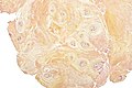
Degenerative disc disease - intermed. mag. (WC)
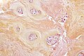
Degenerative disc disease - high mag. (WC)


www:
Sign out
CERVICAL DISC, DISCECTOMY:
- DISC WITH DEGENERATIVE CHANGES.
- NO EVIDENCE OF MALIGNANCY.
LUMBAR DISC, DECOMPRESSION:
- DEGENERATIVE DISC DISEASE.
Schmorl's node
Main article: Schmorl's node
Vertebral fracture
- This deals with benign vertebral fractures.
General
- Usually elderly with osteoporosis.[4]
- Essentially a clinical diagnosis.
- The pathologist's job is to exclude malignancy.
Microscopic
Features:
- Non-viable bone.
DDx:
Sign out
L11 VERTEBRA, BIOPSY:
- BENIGN FRAGMENTS OF BONE.
- BENIGN FIBROCONNECTIVE TISSUE.
- BONE MARROW WITH TRILINEAGE HEMATOPOIESIS.
- NO EVIDENCE OF MALIGNANCY.
See also
References
- ↑ URL: http://www.spineuniverse.com/conditions/synovial-cysts-spine. Accessed on: 5 November 2010.
- ↑ URL: http://www.lab.anhb.uwa.edu.au/mb140/CorePages/Cartilage/Cartil.htm. Accessed on: 2 January 2010.
- ↑ Modic, MT.; Ross, JS. (Oct 2007). "Lumbar degenerative disk disease.". Radiology 245 (1): 43-61. doi:10.1148/radiol.2451051706. PMID 17885180.
- ↑ Sisodia, GB. (2013). "Methods of predicting vertebral body fractures of the lumbar spine.". World J Orthop 4 (4): 241-247. doi:10.5312/wjo.v4.i4.241. PMID 24147259.