Difference between revisions of "Spindle cell"
Jump to navigation
Jump to search
(+images) |
(→Images) |
||
| Line 15: | Line 15: | ||
Image:Urinary bladder muscularis mucosae -- very high mag.jpg | Benign smooth muscle cells of the [[urinary bladder]]. (WC) | Image:Urinary bladder muscularis mucosae -- very high mag.jpg | Benign smooth muscle cells of the [[urinary bladder]]. (WC) | ||
Image:Schwannoma_-_Antoni_A_and_B_-_very_high_mag.jpg | Spindle cells of a schwannoma. (WC) | Image:Schwannoma_-_Antoni_A_and_B_-_very_high_mag.jpg | Spindle cells of a schwannoma. (WC) | ||
</gallery> | |||
====Shapes==== | |||
<gallery> | |||
Image:Drop spindle from Egypt.jpg | A spindle. (WC) | |||
Image:Vesica Piscis.svg | Vesica piscis. (WC) | |||
Image:Ellipse parameters 2.svg | Ellipse. (WC) | |||
</gallery> | </gallery> | ||
Revision as of 05:41, 28 January 2015

Spindle cell is a histomorphologic descriptor used in pathology.
Definition
It refers to a cell that is tapered at both ends.[1]
Notes:
- A taper gradually decreases toward one end [of the cross-section or width].[2]
- Image: Taperred thread (qcfocus.com).
- Spindle cells can have "pointy" ends (typical for nerves) or "rounded" ends (typical for muscle), i.e. be ellipitcal or vesica piscis.
Images
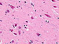
Spindle neurons. (WC)
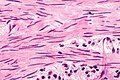
Benign smooth muscle cells of the urinary bladder. (WC)
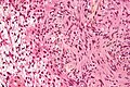
Spindle cells of a schwannoma. (WC)


Shapes

A spindle. (WC)

Vesica piscis. (WC)
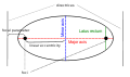
Ellipse. (WC)


See also
References
- ↑ URL: http://www.medterms.com/script/main/art.asp?articlekey=25657. Accessed on: 2 February 2011.
- ↑ URL: http://dictionary.reference.com/browse/taper. Accessed on: 3 February 2011.