Difference between revisions of "Simple endometrial hyperplasia"
Jump to navigation
Jump to search
(redirect w/ cat.) |
|||
| (2 intermediate revisions by the same user not shown) | |||
| Line 1: | Line 1: | ||
{{ Infobox diagnosis | |||
| Name = {{PAGENAME}} | |||
| Image = Simple_endometrial_hyperplasia_-_low_mag.jpg | |||
| Width = | |||
| Caption = Simple endometrial hyperplasia. [[H&E stain]]. | |||
| Synonyms = | |||
| Micro = irregular dilated glands (described as "animal shapes"), variation of gland size, normal gland density (gland area in plane of section/total area ~= 1/3), +/-nuclear atypia (see below) | |||
| Subtypes = with atypia, without atypia | |||
| LMDDx = [[disordered proliferative endometrium]], [[complex endometrial hyperplasia]], [[Atrophic endometrium|cystic atrophy of the endometrium]], [[benign endometrial polyp]] | |||
| Stains = | |||
| IHC = | |||
| EM = | |||
| Molecular = | |||
| IF = | |||
| Gross = | |||
| Grossing = | |||
| Site = [[endometrium]] | |||
| Assdx = | |||
| Syndromes = | |||
| Clinicalhx = | |||
| Signs = [[abnormal uterine bleeding]] | |||
| Symptoms = | |||
| Prevalence = | |||
| Bloodwork = | |||
| Rads = | |||
| Endoscopy = | |||
| Prognosis = good, may progress to [[endometrial carcinoma]] - esp. with atypia | |||
| Other = | |||
| ClinDDx = other cause of [[abnormal uterine bleeding]] | |||
| Tx = | |||
}} | |||
'''Simple endometrial hyperplasia''', abbreviated '''SEH''', is an uncommon pre-malignant change of the [[endometrium]]. Like [[complex endometrial hyperplasia]], it is subdivided into ''with atypia'' and ''without atypia''. | |||
==General== | |||
*More common than simple endometrial hyperplasia with atypia. | |||
*Very low risk for progressing to [[endometrioid endometrial carcinoma]]. | |||
==Microscopic== | |||
Features:<ref name=Ref_GP236>{{Ref GP|236}}</ref> | |||
*Irregular dilated glands (with large lumens) - '''key feature'''. | |||
**Glands described as "animal shapes". | |||
*Variation of gland size. | |||
*Normal gland density (gland area in plane of section/total area ~= 1/3). | |||
*+/-Nuclear atypia:<ref>{{Cite journal | last1 = Silverberg | first1 = SG. | title = Problems in the differential diagnosis of endometrial hyperplasia and carcinoma. | journal = Mod Pathol | volume = 13 | issue = 3 | pages = 309-27 | month = Mar | year = 2000 | doi = 10.1038/modpathol.3880053 | PMID = 10757341 }}</ref> | |||
**Loss of basal nuclear stratification. | |||
**Nuclear size variation. | |||
**Nuclear rounding. | |||
***Nuclei lacking atypical = uniform columnar nuclei. | |||
**Nucleoli. | |||
**Hyperchromasia or [[vesicular nuclei]]. | |||
Notes: | |||
*There are no universally accepted criteria for atypia. Different sources list different features. | |||
*A proposal for atypia (all should be present): | |||
*#Increased NC ratio. | |||
*#*Atypical: ~ 1:2 | |||
*#*Not atypical: ~1:3. | |||
*#Oval nuclei with small major axis to minor axis ratio. | |||
*#*Atypical: major axis:minor axis = <=2:1. | |||
*#*Not atypical: major axis:minor axis = >=3:1 | |||
*#**NB: round nuclei: major axis:minor axis = 1:1. | |||
*#Small nucleoli (~1/5 the size of the nucleus). | |||
DDx: | |||
*[[Disordered proliferative phase]]. | |||
*[[Complex endometrial hyperplasia]] - has increased gland-to-stroma ratio. | |||
*[[Atrophic endometrium|Cystic atrophy of the endometrium]] - does not have proliferative activity.<ref name=pmid16873562>{{Cite journal | last1 = McCluggage | first1 = WG. | title = My approach to the interpretation of endometrial biopsies and curettings. | journal = J Clin Pathol | volume = 59 | issue = 8 | pages = 801-12 | month = Aug | year = 2006 | doi = 10.1136/jcp.2005.029702 | PMID = 16873562 | PMC = 1860448 }}</ref> | |||
*[[Benign endometrial polyp]] - has thick-walled blood vessels; simple endometrial hyperplasia should not be diagnosed in a polyp.<ref name=pmid16873562/> | |||
===Images=== | |||
<gallery> | |||
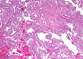
Image:Simple_endometrial_hyperplasia_-_low_mag.jpg | Simple endometrial hyperplasia - low mag. (WC) | |||
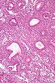
Image:Simple_endometrial_hyperplasia_-_high_mag.jpg | Simple endometrial hyperplasia - high mag. (WC) | |||
</gallery> | |||
www: | |||
*[http://www.webpathology.com/image.asp?n=1&Case=568 Simple endometrial hyperplasia without atypia (webpathology.com)]. | |||
==See also== | |||
*[[Endometrium]]. | |||
*[[Endometrial hyperplasia]]. | |||
==References== | |||
{{Reflist|2}} | |||
[[Category:Endometrium]] | |||
[[Category:Diagnosis]] | [[Category:Diagnosis]] | ||
Latest revision as of 15:47, 1 June 2015
| Simple endometrial hyperplasia | |
|---|---|
| Diagnosis in short | |
 Simple endometrial hyperplasia. H&E stain. | |
|
| |
| LM | irregular dilated glands (described as "animal shapes"), variation of gland size, normal gland density (gland area in plane of section/total area ~= 1/3), +/-nuclear atypia (see below) |
| Subtypes | with atypia, without atypia |
| LM DDx | disordered proliferative endometrium, complex endometrial hyperplasia, cystic atrophy of the endometrium, benign endometrial polyp |
| Site | endometrium |
|
| |
| Signs | abnormal uterine bleeding |
| Prognosis | good, may progress to endometrial carcinoma - esp. with atypia |
| Clin. DDx | other cause of abnormal uterine bleeding |
Simple endometrial hyperplasia, abbreviated SEH, is an uncommon pre-malignant change of the endometrium. Like complex endometrial hyperplasia, it is subdivided into with atypia and without atypia.
General
- More common than simple endometrial hyperplasia with atypia.
- Very low risk for progressing to endometrioid endometrial carcinoma.
Microscopic
Features:[1]
- Irregular dilated glands (with large lumens) - key feature.
- Glands described as "animal shapes".
- Variation of gland size.
- Normal gland density (gland area in plane of section/total area ~= 1/3).
- +/-Nuclear atypia:[2]
- Loss of basal nuclear stratification.
- Nuclear size variation.
- Nuclear rounding.
- Nuclei lacking atypical = uniform columnar nuclei.
- Nucleoli.
- Hyperchromasia or vesicular nuclei.
Notes:
- There are no universally accepted criteria for atypia. Different sources list different features.
- A proposal for atypia (all should be present):
- Increased NC ratio.
- Atypical: ~ 1:2
- Not atypical: ~1:3.
- Oval nuclei with small major axis to minor axis ratio.
- Atypical: major axis:minor axis = <=2:1.
- Not atypical: major axis:minor axis = >=3:1
- NB: round nuclei: major axis:minor axis = 1:1.
- Small nucleoli (~1/5 the size of the nucleus).
- Increased NC ratio.
DDx:
- Disordered proliferative phase.
- Complex endometrial hyperplasia - has increased gland-to-stroma ratio.
- Cystic atrophy of the endometrium - does not have proliferative activity.[3]
- Benign endometrial polyp - has thick-walled blood vessels; simple endometrial hyperplasia should not be diagnosed in a polyp.[3]
Images
Simple endometrial hyperplasia - low mag. (WC)
Simple endometrial hyperplasia - high mag. (WC)

www:
See also
References
- ↑ Nucci, Marisa R.; Oliva, Esther (2009). Gynecologic Pathology: A Volume in Foundations in Diagnostic Pathology Series (1st ed.). Churchill Livingstone. pp. 236. ISBN 978-0443069208.
- ↑ Silverberg, SG. (Mar 2000). "Problems in the differential diagnosis of endometrial hyperplasia and carcinoma.". Mod Pathol 13 (3): 309-27. doi:10.1038/modpathol.3880053. PMID 10757341.
- ↑ 3.0 3.1 McCluggage, WG. (Aug 2006). "My approach to the interpretation of endometrial biopsies and curettings.". J Clin Pathol 59 (8): 801-12. doi:10.1136/jcp.2005.029702. PMC 1860448. PMID 16873562. https://www.ncbi.nlm.nih.gov/pmc/articles/PMC1860448/.