Difference between revisions of "Rhabdomyosarcoma"
(more) |
|||
| (41 intermediate revisions by the same user not shown) | |||
| Line 1: | Line 1: | ||
'''Rhabdomyosarcoma''', often abbreviated ''RMS'', is a malignant tumour of skeletal muscle. | {{ Infobox diagnosis | ||
| Name = {{PAGENAME}} | |||
| Image = Alveolar_rhabdomyosarcoma_-_very_high_mag.jpg | |||
| Width = | |||
| Caption = Alveolar rhabdomyosarcoma. [[H&E stain]]. | |||
| Synonyms = | |||
| Micro = +/-rhabdomyoblasts (eccentric nucleus, moderate amount of intensly eosinophilic cytoplasm, striations - not common); alveolar RMS: alveolus-like pattern (classic); embryonal RMS: [[small round cell tumour]] | |||
| Subtypes = embryonal (spindle cell subtype, botryoid), alveolar (translocation-positive, translocation-negative), undifferentiated | |||
| LMDDx = [[small round cell tumours]] - esp. [[small cell carcinoma]] and (large cell) [[lymphoma]]s | |||
| Stains = | |||
| IHC = desmin (best marker) +ve, actin +ve, myogenin +ve, CD56 +ve (common), synaptophysin -ve/+ve, chromogranin -ve/+ve, cytokeratins -ve/+ve | |||
| EM = sarcomeric like structures - typically in U-shaped cells | |||
| Molecular = alveolar RMS (~85% of cases): t(2,13) PAX3/FKHR fusion gene ''or'' t(1,13) PAX7/FKHR fusion gene | |||
| IF = | |||
| Gross = | |||
| Grossing = | |||
| Site = [[soft tissue]] - skeletal muscle site (alveolar RMS), non-skeletal muscle site (embryonal RMS) | |||
| Assdx = | |||
| Syndromes = [[DICER1 syndrome]] for ''embryonal rhabdomyosarcoma'' | |||
| Clinicalhx = alveolar RMS: young adult or adolescent; embryonal RMS: typically <10 years old | |||
| Signs = | |||
| Symptoms = | |||
| Prevalence = not common | |||
| Bloodwork = | |||
| Rads = | |||
| Endoscopy = | |||
| Prognosis = | |||
| Other = | |||
| ClinDDx = other soft tissue tumours | |||
| Tx = | |||
}} | |||
'''Rhabdomyosarcoma''', often abbreviated '''RMS''', is a [[malignant]] tumour of skeletal muscle. | |||
==General== | ==General== | ||
*Most common paediatric sarcoma. | *Most common paediatric [[sarcoma]]. | ||
**Classically in the head and neck region.<ref name=pmid10465231>{{Cite journal | last1 = Rosenthal | first1 = TC. | last2 = Kraybill | first2 = W. | title = Soft tissue sarcomas: integrating primary care recognition with tertiary care center treatment. | journal = Am Fam Physician | volume = 60 | issue = 2 | pages = 567-72 | month = Aug | year = 1999 | doi = | PMID = 10465231 | URL = http://www.aafp.org/afp/1999/0801/p567.html }}</ref> | |||
*Most common sarcoma in [[Li-Fraumeni syndrome]].<ref name=PST14feb11>Thorner, Paul S. 14 February 2011.</ref> | |||
*~6% of all childhood cancer. | *~6% of all childhood cancer. | ||
| Line 10: | Line 43: | ||
#*Usually young adults/adolescents. | #*Usually young adults/adolescents. | ||
#*Early mets common. | #*Early mets common. | ||
#* | #*Usually arises in regions with skeletal muscle. | ||
#Embryonal rhabdomyosarcoma. | #Embryonal rhabdomyosarcoma. | ||
#*Usual <10 years old. | #*Usual <10 years old. | ||
#*Typically locally invasive. | #*Typically locally invasive. | ||
#* | #*Usually arises in regions '''without''' skeletal muscle. | ||
Less common types:<ref name=pmid12110339>{{Cite journal | last1 = Hicks | first1 = J. | last2 = Flaitz | first2 = C. | title = Rhabdomyosarcoma of the head and neck in children. | journal = Oral Oncol | volume = 38 | issue = 5 | pages = 450-9 | month = Jul | year = 2002 | doi = | PMID = 12110339 }}</ref> | |||
#Undifferentiated rhabdomyosarcoma. | |||
#Botryoid - may be considered a subtype of embryonal RMS. | |||
#Spindle cell - may be considered a subtype of embryonal RMS. | |||
Notes: | |||
*How to remember the special types ''BUS'': '''b'''otryoid, '''u'''ndifferentiated, '''s'''pindle. | |||
*The above is the international classification. Several classification of RMS exist - see: [http://www.nature.com/modpathol/journal/v14/n5/fig_tab/3880339t1.html#figure-title Classifications of Rhabdomyosarcoma].<ref name=pmid11353062>{{Cite journal | last1 = Parham | first1 = DM. | title = Pathologic classification of rhabdomyosarcomas and correlations with molecular studies. | journal = Mod Pathol | volume = 14 | issue = 5 | pages = 506-14 | month = May | year = 2001 | doi = 10.1038/modpathol.3880339 | PMID = 11353062 }} | |||
</ref> | |||
====Molecular and histologic==== | ====Molecular and histologic==== | ||
| Line 22: | Line 65: | ||
Notes: | Notes: | ||
*Translocation-negative alveolar RMS shares gene expression | *Translocation-negative alveolar RMS shares gene expression profiling characteristics with embryonal RMS -- suggesting these can be grouped together. | ||
==Gross== | |||
Sarcoma botryoides (embryonal RMS) - distinctive appearance: | |||
*Grapes on the vine-like clusters. | |||
**Found in [[urinary bladder]], [[vagina]]. | |||
Image: | |||
*[http://www.flickr.com/photos/35441329@N05/4985992462/in/photostream/ Sarcoma Boytriodes (flickr.com)]. | |||
==Microscopic== | ==Microscopic== | ||
===Alveolar rhabdomyosarcoma=== | ===Alveolar rhabdomyosarcoma=== | ||
Features:<ref name=PST14feb11 | Features:<ref name=PST14feb11/> | ||
*Alveolus-like pattern -- '''key low-power feature'''. | *Alveolus-like pattern -- '''key low-power feature'''. | ||
**Fibrous septae lined by tumour cells. | **Fibrous septae lined by tumour cells. | ||
| Line 37: | Line 88: | ||
Other features: | Other features: | ||
*Nuclear pleomorphism - common. | *[[Nuclear pleomorphism]] - common. | ||
*Mitoses - common. | *Mitoses - common. | ||
Notes: | Notes: | ||
*Well-differentiated rhabdomyoblasts are uncommon in alveolar RMS. | *Well-differentiated rhabdomyoblasts are uncommon in alveolar RMS. | ||
DDx: | |||
*[[Alveolar soft part sarcoma]]. | |||
*Skeletal muscle regeneration.<ref name=pmid9762546>{{Cite journal | last1 = Guillou | first1 = L. | last2 = Coquet | first2 = M. | last3 = Chaubert | first3 = P. | last4 = Coindre | first4 = JM. | title = Skeletal muscle regeneration mimicking rhabdomyosarcoma: a potential diagnostic pitfall. | journal = Histopathology | volume = 33 | issue = 2 | pages = 136-44 | month = Aug | year = 1998 | doi = | PMID = 9762546 }}</ref> | |||
====Images==== | |||
<gallery> | |||
Image:Alveolar_rhabdomyosarcoma_-_intermed_mag.jpg | Alveolar RMS - intermed. mag. (WC) | |||
Image:Alveolar_rhabdomyosarcoma_-_very_high_mag.jpg | Alveolar RMS - very high mag. (WC) | |||
</gallery> | |||
www: | |||
*[http://path.upmc.edu/cases/case489.html Alveolar RMS - several images (upmc.edu)]. | |||
===Embryonal rhabdomyosarcoma=== | ===Embryonal rhabdomyosarcoma=== | ||
Features:<ref name=PST14feb11 | Features:<ref name=PST14feb11/> | ||
*Randomly arranged small cells. | *Randomly arranged small cells. | ||
*Myxoid matrix. | *[[Myxoid]] matrix. | ||
*Strap cells: | *Strap cells: | ||
**Tadpole-like morphology. | **Tadpole-like morphology. | ||
| Line 53: | Line 116: | ||
**Moderate amount of intensly eosinophilic cytoplasm. | **Moderate amount of intensly eosinophilic cytoplasm. | ||
**Striations -- if you're really lucky; these are not common. | **Striations -- if you're really lucky; these are not common. | ||
DDx: | |||
*[[Small round cell tumours]]. | |||
*[[Pleuropulmonary blastoma]].<ref name=pmid20677658>{{Cite journal | last1 = Chen | first1 = S. | last2 = Wang | first2 = S. | last3 = Gao | first3 = J. | last4 = Zhang | first4 = S. | title = [Pleuropulmonary blastoma: a clinicopathological analysis]. | journal = Zhongguo Fei Ai Za Zhi | volume = 13 | issue = 5 | pages = 550-3 | month = May | year = 2010 | doi = 10.3779/j.issn.1009-3419.2010.05.31 | PMID = 20677658 }}</ref> | |||
Images: | |||
*[http://path.upmc.edu/cases/case204/micro.html Embryonal RMS - several images (upmc.edu)]. | |||
====Subtypes of embryonal RMS==== | ====Subtypes of embryonal RMS==== | ||
| Line 58: | Line 128: | ||
Common subtypes: | Common subtypes: | ||
#Botryoid subtype: | #Botryoid subtype ([[AKA]] ''sarcoma botryoides''): | ||
#*Gross: Grape-like morphology. | #*Gross: Grape-like morphology. | ||
#*Microscopic: Non-proliferating layer deep to the surface (" | #*Microscopic: Non-proliferating layer deep to the surface ("Cambium layer"). | ||
#Spindle cell subtype. | #Spindle cell subtype. | ||
#*General: may mimic [[leiomyosarcoma]] -- which is not common in the pediatric population. | #*General: may mimic [[leiomyosarcoma]] (complete with [[vesicular pattern]]) -- which is not common in the pediatric population. | ||
#*Microscopic: vesicular growth pattern, spindle cells. | #*Microscopic: vesicular growth pattern, spindle cells. | ||
Notes: | |||
*Cambium layer = cellular region deep to epithelial component.<ref>URL: [http://www.medilexicon.com/medicaldictionary.php?t=48297 http://www.medilexicon.com/medicaldictionary.php?t=48297]. Accessed on: 9 August 2011.</ref> | |||
**Can be thought of as the opposite of a "Grenz zone" -- which is a paucicellular zone between tumour and epithelium. | |||
===Anaplasia=== | |||
Criteria: | |||
#Hyperchromatic nuclei with size variation greater or equal to 3x. | |||
#Multipolar (atypical) mitotic figures. | |||
Subclassification: | |||
#Focal - a few cells. | |||
#Diffuse - cluster or sheets of anaplasia. | |||
Notes: | |||
*Not subtle - can identify at low power. | |||
*Seen in 10-15% of RMS. | |||
**More common in older individuals. | |||
*Poorer prognosis in embryonal RMS. | |||
**No change in prognosis in alveolar RMS. | |||
==IHC== | ==IHC== | ||
| Line 71: | Line 161: | ||
*Myogenin. | *Myogenin. | ||
==[[ | For [[head and neck pathology|head and neck]] RMS:<ref name=pmid18487991>{{Cite journal | last1 = Bahrami | first1 = A. | last2 = Gown | first2 = AM. | last3 = Baird | first3 = GS. | last4 = Hicks | first4 = MJ. | last5 = Folpe | first5 = AL. | title = Aberrant expression of epithelial and neuroendocrine markers in alveolar rhabdomyosarcoma: a potentially serious diagnostic pitfall. | journal = Mod Pathol | volume = 21 | issue = 7 | pages = 795-806 | month = Jul | year = 2008 | doi = 10.1038/modpathol.2008.86 | PMID = 18487991 }}</ref> | ||
*CD56 +ve. | |||
*Synaptophysin -ve/+ve (seen in 12 of 37 cases<ref name=pmid18487991/>). | |||
*Chromogranin A -ve/+ve (seen in 8 of 36 cases<ref name=pmid18487991/>). | |||
*Wide-spectrum cytokeratin -ve/+ve. | |||
*CAM5.2 -ve/+ve. | |||
For [[urinary bladder]] RMS in adults: | |||
*Myogenin +ve. | |||
*Desmin +ve. | |||
*Keratins -ve.<ref name=pmid21762516>{{Cite journal | last1 = Bing | first1 = Z. | last2 = Zhang | first2 = PJ. | title = Adult urinary bladder tumors with rhabdomyosarcomatous differentiation: clinical, pathological and immunohistochemical studies. | journal = Diagn Pathol | volume = 6 | issue = | pages = 66 | month = | year = 2011 | doi = 10.1186/1746-1596-6-66 | PMID = 21762516 }}</ref> | |||
**Keratin positive tumours are considered ''rhabdomyosarcomatous sarcomatoid carcinoma'' or ''sarcomatoid carcinoma with rhabdomyosarcomatous differentiation''. | |||
===Subtyping via IHC=== | |||
PST proposes<ref name=PST14feb11/> the following (presumably based on Makawitz et al.<ref name=pmid18788888>{{cite journal |author=Makawita S, Ho M, Durbin AD, Thorner PS, Malkin D, Somers GR |title=Expression of insulin-like growth factor pathway proteins in rhabdomyosarcoma: IGF-2 expression is associated with translocation-negative tumors |journal=Pediatr. Dev. Pathol. |volume=12 |issue=2 |pages=127–35 |year=2009 |pmid=18788888 |doi=10.2350/08-05-0477.1 |url=}}</ref>): | |||
{| class="wikitable sortable" style="margin-left:auto;margin-right:auto" | |||
| '''IHC''' | |||
| '''Translocation positive<br> alveolar RMS | |||
| '''Embryonal RMS''' | |||
| '''Translocation negative<br> alveolar RMS''' | |||
|- | |||
| myogenin | |||
| +ve -- diffuse | |||
| +ve -- focal | |||
| +ve -- diffuse | |||
|- | |||
| EGFR | |||
| -ve | |||
| +ve | |||
| -ve | |||
|- | |||
| P-cadherin | |||
| +ve | |||
| -ve | |||
| -ve | |||
|- | |||
| IGF2 | |||
| -ve | |||
| +ve | |||
| +ve | |||
|} | |||
A paper by Wachtel at al.<ref>{{cite journal |author=Wachtel M, Runge T, Leuschner I, ''et al.'' |title=Subtype and prognostic classification of rhabdomyosarcoma by immunohistochemistry |journal=J. Clin. Oncol. |volume=24 |issue=5 |pages=816–22 |year=2006 |month=February |pmid=16391296 |doi=10.1200/JCO.2005.03.4934 |url=}}</ref> proposes the use of: | |||
* ''AP2beta'' and ''P-cadherin'' +ve in translocation positive alveolar RMS, and | |||
* ''EGFR'' and ''fibrillin-2'' +ve in embryonal RMS and translocation negative alveolar RMS. | |||
==[[Electron microscopy]]== | |||
Features: | Features: | ||
*Sarcomeric like structures - | *Sarcomeric like structures - usually in "bent" cells; cells that are U-shaped. | ||
==Molecular diagnostics== | ==Molecular diagnostics== | ||
===Alveolar rhabdomyosarcoma=== | ===Alveolar rhabdomyosarcoma=== | ||
Common translocations (~ | Common translocations (~85% of cases): | ||
*t(1,13). | *t(1,13). | ||
**PAX7/FKHR fusion gene. | **PAX7/FKHR fusion gene. | ||
**Seen in approx. 15% of cases. | |||
*t(2,13).<ref>URL: [http://www.ncbi.nlm.nih.gov/omim/606597 http://www.ncbi.nlm.nih.gov/omim/606597]. Accessed on: 18 August 2010.</ref> | *t(2,13).<ref>URL: [http://www.ncbi.nlm.nih.gov/omim/606597 http://www.ncbi.nlm.nih.gov/omim/606597]. Accessed on: 18 August 2010.</ref> | ||
**PAX3/FKHR fusion gene. | **PAX3/FKHR fusion gene. | ||
**Seen in approx. 70% of cases. | |||
Notes: | Notes: | ||
*t(1,13) vs. t(2,13) -- t(1,13) usually: younger age, extremity lesion, localized disease, better survival. | *t(1,13) vs. t(2,13) -- t(1,13) usually: younger age, extremity lesion, localized disease, better survival. | ||
*Several uncommon [[translocations]] exist. | *Several uncommon [[translocations]] exist. | ||
*'''Important''' for [[urinary bladder]] lesions in adults: the presence of a translocation is more-or-less required for the diagnosis of RMS.<ref name=pmid21762516>{{Cite journal | last1 = Bing | first1 = Z. | last2 = Zhang | first2 = PJ. | title = Adult urinary bladder tumors with rhabdomyosarcomatous differentiation: clinical, pathological and immunohistochemical studies. | journal = Diagn Pathol | volume = 6 | issue = | pages = 66 | month = | year = 2011 | doi = 10.1186/1746-1596-6-66 | PMID = 21762516 }}</ref> | |||
**It is suggested that keratin negative tumours without molecular testing to corroborate the impression of RMS be referred to as ''rhabdomyomatous tumours''.<ref name=pmid21762516/> | |||
===Embryonal rhabdomyosarcoma=== | |||
*Chromosome 11p loss of heterozygosity.<ref name=pmid17652054>{{Cite journal | last1 = Gallego Melcón | first1 = S. | last2 = Sánchez de Toledo Codina | first2 = J. | title = Molecular biology of rhabdomyosarcoma. | journal = Clin Transl Oncol | volume = 9 | issue = 7 | pages = 415-9 | month = Jul | year = 2007 | doi = | PMID = 17652054 }}</ref> | |||
Note: | |||
*Not used for diagnosis. | |||
==See also== | ==See also== | ||
Latest revision as of 12:40, 24 March 2024
| Rhabdomyosarcoma | |
|---|---|
| Diagnosis in short | |
 Alveolar rhabdomyosarcoma. H&E stain. | |
|
| |
| LM | +/-rhabdomyoblasts (eccentric nucleus, moderate amount of intensly eosinophilic cytoplasm, striations - not common); alveolar RMS: alveolus-like pattern (classic); embryonal RMS: small round cell tumour |
| Subtypes | embryonal (spindle cell subtype, botryoid), alveolar (translocation-positive, translocation-negative), undifferentiated |
| LM DDx | small round cell tumours - esp. small cell carcinoma and (large cell) lymphomas |
| IHC | desmin (best marker) +ve, actin +ve, myogenin +ve, CD56 +ve (common), synaptophysin -ve/+ve, chromogranin -ve/+ve, cytokeratins -ve/+ve |
| EM | sarcomeric like structures - typically in U-shaped cells |
| Molecular | alveolar RMS (~85% of cases): t(2,13) PAX3/FKHR fusion gene or t(1,13) PAX7/FKHR fusion gene |
| Site | soft tissue - skeletal muscle site (alveolar RMS), non-skeletal muscle site (embryonal RMS) |
|
| |
| Syndromes | DICER1 syndrome for embryonal rhabdomyosarcoma |
|
| |
| Clinical history | alveolar RMS: young adult or adolescent; embryonal RMS: typically <10 years old |
| Prevalence | not common |
| Clin. DDx | other soft tissue tumours |
Rhabdomyosarcoma, often abbreviated RMS, is a malignant tumour of skeletal muscle.
General
- Most common paediatric sarcoma.
- Classically in the head and neck region.[1]
- Most common sarcoma in Li-Fraumeni syndrome.[2]
- ~6% of all childhood cancer.
Classification
Histologic
- Alveolar rhabdomyosarcoma.
- Usually young adults/adolescents.
- Early mets common.
- Usually arises in regions with skeletal muscle.
- Embryonal rhabdomyosarcoma.
- Usual <10 years old.
- Typically locally invasive.
- Usually arises in regions without skeletal muscle.
Less common types:[3]
- Undifferentiated rhabdomyosarcoma.
- Botryoid - may be considered a subtype of embryonal RMS.
- Spindle cell - may be considered a subtype of embryonal RMS.
Notes:
- How to remember the special types BUS: botryoid, undifferentiated, spindle.
- The above is the international classification. Several classification of RMS exist - see: Classifications of Rhabdomyosarcoma.[4]
Molecular and histologic
- Translocation-positive alveolar RMS.
- Translocation-negative alveolar RMS.
- Embryonal RMS.
Notes:
- Translocation-negative alveolar RMS shares gene expression profiling characteristics with embryonal RMS -- suggesting these can be grouped together.
Gross
Sarcoma botryoides (embryonal RMS) - distinctive appearance:
- Grapes on the vine-like clusters.
- Found in urinary bladder, vagina.
Image:
Microscopic
Alveolar rhabdomyosarcoma
Features:[2]
- Alveolus-like pattern -- key low-power feature.
- Fibrous septae lined by tumour cells.
- Cells may "fall-off" the septa, i.e. be detached/scattered in the alveolus-like space.
- Space between fibrous sepate may be filled with tumour = solid variant of alveolar rhabdomyosarcoma.
- Fibrous septae lined by tumour cells.
- Rhabdomyoblasts - essentially diagnostic.
- Eccentric nucleus.
- Moderate amount of intensly eosinophilic cytoplasm.
- Striations -- if you're really lucky; these are not common.
Other features:
- Nuclear pleomorphism - common.
- Mitoses - common.
Notes:
- Well-differentiated rhabdomyoblasts are uncommon in alveolar RMS.
DDx:
- Alveolar soft part sarcoma.
- Skeletal muscle regeneration.[5]
Images
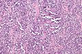
Alveolar RMS - intermed. mag. (WC)
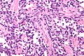
Alveolar RMS - very high mag. (WC)

www:
Embryonal rhabdomyosarcoma
Features:[2]
- Randomly arranged small cells.
- Myxoid matrix.
- Strap cells:
- Tadpole-like morphology.
- Rhabdomyoblasts - essentially diagnostic.
- Eccentric nucleus.
- Moderate amount of intensly eosinophilic cytoplasm.
- Striations -- if you're really lucky; these are not common.
DDx:
Images:
Subtypes of embryonal RMS
There are two common subtypes of embryonal RMS. Both of them have a better prognosis that embryonal RMS not otherwise specified (NOS).
Common subtypes:
- Botryoid subtype (AKA sarcoma botryoides):
- Gross: Grape-like morphology.
- Microscopic: Non-proliferating layer deep to the surface ("Cambium layer").
- Spindle cell subtype.
- General: may mimic leiomyosarcoma (complete with vesicular pattern) -- which is not common in the pediatric population.
- Microscopic: vesicular growth pattern, spindle cells.
Notes:
- Cambium layer = cellular region deep to epithelial component.[7]
- Can be thought of as the opposite of a "Grenz zone" -- which is a paucicellular zone between tumour and epithelium.
Anaplasia
Criteria:
- Hyperchromatic nuclei with size variation greater or equal to 3x.
- Multipolar (atypical) mitotic figures.
Subclassification:
- Focal - a few cells.
- Diffuse - cluster or sheets of anaplasia.
Notes:
- Not subtle - can identify at low power.
- Seen in 10-15% of RMS.
- More common in older individuals.
- Poorer prognosis in embryonal RMS.
- No change in prognosis in alveolar RMS.
IHC
Panel of muscle markers -- DAM:
- Desmin (best marker).
- Actin.
- Myogenin.
For head and neck RMS:[8]
- CD56 +ve.
- Synaptophysin -ve/+ve (seen in 12 of 37 cases[8]).
- Chromogranin A -ve/+ve (seen in 8 of 36 cases[8]).
- Wide-spectrum cytokeratin -ve/+ve.
- CAM5.2 -ve/+ve.
For urinary bladder RMS in adults:
- Myogenin +ve.
- Desmin +ve.
- Keratins -ve.[9]
- Keratin positive tumours are considered rhabdomyosarcomatous sarcomatoid carcinoma or sarcomatoid carcinoma with rhabdomyosarcomatous differentiation.
Subtyping via IHC
PST proposes[2] the following (presumably based on Makawitz et al.[10]):
| IHC | Translocation positive alveolar RMS |
Embryonal RMS | Translocation negative alveolar RMS |
| myogenin | +ve -- diffuse | +ve -- focal | +ve -- diffuse |
| EGFR | -ve | +ve | -ve |
| P-cadherin | +ve | -ve | -ve |
| IGF2 | -ve | +ve | +ve |
A paper by Wachtel at al.[11] proposes the use of:
- AP2beta and P-cadherin +ve in translocation positive alveolar RMS, and
- EGFR and fibrillin-2 +ve in embryonal RMS and translocation negative alveolar RMS.
Electron microscopy
Features:
- Sarcomeric like structures - usually in "bent" cells; cells that are U-shaped.
Molecular diagnostics
Alveolar rhabdomyosarcoma
Common translocations (~85% of cases):
- t(1,13).
- PAX7/FKHR fusion gene.
- Seen in approx. 15% of cases.
- t(2,13).[12]
- PAX3/FKHR fusion gene.
- Seen in approx. 70% of cases.
Notes:
- t(1,13) vs. t(2,13) -- t(1,13) usually: younger age, extremity lesion, localized disease, better survival.
- Several uncommon translocations exist.
- Important for urinary bladder lesions in adults: the presence of a translocation is more-or-less required for the diagnosis of RMS.[9]
- It is suggested that keratin negative tumours without molecular testing to corroborate the impression of RMS be referred to as rhabdomyomatous tumours.[9]
Embryonal rhabdomyosarcoma
- Chromosome 11p loss of heterozygosity.[13]
Note:
- Not used for diagnosis.
See also
References
- ↑ Rosenthal, TC.; Kraybill, W. (Aug 1999). "Soft tissue sarcomas: integrating primary care recognition with tertiary care center treatment.". Am Fam Physician 60 (2): 567-72. PMID 10465231.
- ↑ 2.0 2.1 2.2 2.3 Thorner, Paul S. 14 February 2011.
- ↑ Hicks, J.; Flaitz, C. (Jul 2002). "Rhabdomyosarcoma of the head and neck in children.". Oral Oncol 38 (5): 450-9. PMID 12110339.
- ↑ Parham, DM. (May 2001). "Pathologic classification of rhabdomyosarcomas and correlations with molecular studies.". Mod Pathol 14 (5): 506-14. doi:10.1038/modpathol.3880339. PMID 11353062.
- ↑ Guillou, L.; Coquet, M.; Chaubert, P.; Coindre, JM. (Aug 1998). "Skeletal muscle regeneration mimicking rhabdomyosarcoma: a potential diagnostic pitfall.". Histopathology 33 (2): 136-44. PMID 9762546.
- ↑ Chen, S.; Wang, S.; Gao, J.; Zhang, S. (May 2010). "[Pleuropulmonary blastoma: a clinicopathological analysis].". Zhongguo Fei Ai Za Zhi 13 (5): 550-3. doi:10.3779/j.issn.1009-3419.2010.05.31. PMID 20677658.
- ↑ URL: http://www.medilexicon.com/medicaldictionary.php?t=48297. Accessed on: 9 August 2011.
- ↑ 8.0 8.1 8.2 Bahrami, A.; Gown, AM.; Baird, GS.; Hicks, MJ.; Folpe, AL. (Jul 2008). "Aberrant expression of epithelial and neuroendocrine markers in alveolar rhabdomyosarcoma: a potentially serious diagnostic pitfall.". Mod Pathol 21 (7): 795-806. doi:10.1038/modpathol.2008.86. PMID 18487991.
- ↑ 9.0 9.1 9.2 Bing, Z.; Zhang, PJ. (2011). "Adult urinary bladder tumors with rhabdomyosarcomatous differentiation: clinical, pathological and immunohistochemical studies.". Diagn Pathol 6: 66. doi:10.1186/1746-1596-6-66. PMID 21762516.
- ↑ Makawita S, Ho M, Durbin AD, Thorner PS, Malkin D, Somers GR (2009). "Expression of insulin-like growth factor pathway proteins in rhabdomyosarcoma: IGF-2 expression is associated with translocation-negative tumors". Pediatr. Dev. Pathol. 12 (2): 127–35. doi:10.2350/08-05-0477.1. PMID 18788888.
- ↑ Wachtel M, Runge T, Leuschner I, et al. (February 2006). "Subtype and prognostic classification of rhabdomyosarcoma by immunohistochemistry". J. Clin. Oncol. 24 (5): 816–22. doi:10.1200/JCO.2005.03.4934. PMID 16391296.
- ↑ URL: http://www.ncbi.nlm.nih.gov/omim/606597. Accessed on: 18 August 2010.
- ↑ Gallego Melcón, S.; Sánchez de Toledo Codina, J. (Jul 2007). "Molecular biology of rhabdomyosarcoma.". Clin Transl Oncol 9 (7): 415-9. PMID 17652054.