Difference between revisions of "Rhabdomyoma"
Jump to navigation
Jump to search
| Line 57: | Line 57: | ||
[[Category:Soft tissue lesions]] | [[Category:Soft tissue lesions]] | ||
[[Category:Diagnosis]] | [[Category:Diagnosis]] | ||
[[Category:Head and neck pathology]] | |||
Revision as of 14:32, 4 September 2017
Rhabdomyoma a benign muscle tumour. Often seen in the context of tuberous sclerosis.
General
- May be seen in the context of tuberous sclerosis.
Gross
- Solid, white/tan colour.
Image:
Microscopic
Features - cardiac:[1]
- Spider cells:
- Large polygonal cells (~10-20x RBC diameter):
- Abundant cytoplasm filled with glycogen.
- Large polygonal cells (~10-20x RBC diameter):
Note:
- Fetal rhabdomyomas may have pseudoepitheliomatous hyperplasia.[2]
DDx:
Images
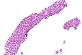
Rhabdomyoma - low mag. (WC)
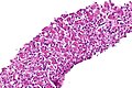
Rhabdomyoma - intermed. mag. (WC)
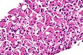
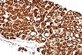
Rhabdomyoma - high mag. (WC)
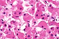
Rhabdomyoma - very high mag. (WC)
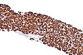
Rhabdomyoma - desmin - intermed. mag. (WC)
Rhabdomyoma - desmin - high mag. (WC)


www
IHC
Features:[2]
- Desmin +ve.
- Myogoblin +ve.
- Actin +ve.
- S-100 -ve.
- Positive in granular cell tumour and hiberoma.
See also
References
- ↑ URL: http://www.brown.edu/Courses/Digital_Path/systemic_path/cardio/rhabdomyoma.html. Accessed on: 19 October 2011.
- ↑ 2.0 2.1 Hansen, T.; Katenkamp, D. (Nov 2005). "Rhabdomyoma of the head and neck: morphology and differential diagnosis.". Virchows Arch 447 (5): 849-54. doi:10.1007/s00428-005-0038-8. PMID 16133368.