Difference between revisions of "Pulmonary embolism"
Jump to navigation
Jump to search
(modify) |
|||
| (40 intermediate revisions by the same user not shown) | |||
| Line 1: | Line 1: | ||
'''Pulmonary embolism''', abbreviated '''PE''', is often on the differential in [[autopsy|autopsies]], as it is not easy to diagnose clinically. | '''Pulmonary embolism''', abbreviated '''PE''', is often on the differential in [[autopsy|autopsies]], as it is not easy to diagnose clinically. ''Pulmonary embolism'' is a non-specific term; it may refer to a number of things, including: | ||
*Pulmonary venous thromboembolism. | |||
*Pulmonary fat embolism. | |||
*Pulmonary foreign body embolism. | |||
*Pulmonary septic embolism. | |||
*Pulmonary bone marrow embolism. | |||
*Pulmonary tumour embolism. | |||
*Pulmonary amniotic fluid embolism. | |||
==Clinical== | PE usually refers to '''pulmonary venous thromboembolism''', abbreviated '''VTE''', if not otherwise specified. | ||
*Shortness of breath (dyspnea) - classic symptom. | |||
==General== | |||
*Relatively uncommon ~ 1 in 1000 adults per year.<ref name=pmid20852464>{{Cite journal | last1 = Meetoo | first1 = D. | title = In too deep: understanding, detecting and managing DVT. | journal = Br J Nurs | volume = 19 | issue = 16 | pages = 1021-7 | month = | year = | doi = | PMID = 20852464 }}</ref> | |||
*Diagnosis in life dependent on strong clinical suspicion and radiology. | |||
===Clinical=== | |||
*Shortness of breath ([[dyspnea]]) - classic symptom. | |||
*Tachycardia. | *Tachycardia. | ||
*Chest pain. | *Chest pain. | ||
*Findings associated with deep vein thrombosis | *Findings associated with deep vein [[thrombosis]]. | ||
**Leg pain. | **Leg pain. | ||
**Leg swelling. | **Leg swelling. | ||
| Line 12: | Line 25: | ||
*Venous thrombosis OR~=12 for PE.<ref name=pmid20648405>{{cite journal |author=Reissig A, Haase U, Schulze E, Lehmann T, Kroegel C |title=[Diagnosis and therapy of pulmonary embolism prior to death] |language=German |journal=Dtsch. Med. Wochenschr. |volume=135 |issue=30 |pages=1477–83 |year=2010 |month=July |pmid=20648405 |doi=10.1055/s-0030-1262435 |url=}}</ref> | *Venous thrombosis OR~=12 for PE.<ref name=pmid20648405>{{cite journal |author=Reissig A, Haase U, Schulze E, Lehmann T, Kroegel C |title=[Diagnosis and therapy of pulmonary embolism prior to death] |language=German |journal=Dtsch. Med. Wochenschr. |volume=135 |issue=30 |pages=1477–83 |year=2010 |month=July |pmid=20648405 |doi=10.1055/s-0030-1262435 |url=}}</ref> | ||
== | ===Mechanism=== | ||
The classic factors are given by ''Virchow's triad'':<ref name=pmid22345594>{{Cite journal | last1 = Reitsma | first1 = PH. | last2 = Versteeg | first2 = HH. | last3 = Middeldorp | first3 = S. | title = Mechanistic view of risk factors for venous thromboembolism. | journal = Arterioscler Thromb Vasc Biol | volume = 32 | issue = 3 | pages = 563-8 | month = Mar | year = 2012 | doi = 10.1161/ATVBAHA.111.242818 | PMID = 22345594 }}</ref><ref name=pmid20852464>{{Cite journal | last1 = Meetoo | first1 = D. | title = In too deep: understanding, detecting and managing DVT. | journal = Br J Nurs | volume = 19 | issue = 16 | pages = 1021-7 | month = | year = | doi = | PMID = 20852464 }}</ref> | |||
#Hypercoagulability. | |||
#Endothelial dysfunction/injury. | |||
#Stasis. | |||
==Gross== | Note: | ||
*Intravascular | *The triad has a limited practical use. Like many questions about mechanism, the greatest utility, might be pimping medical students and residents. | ||
===Risks factors venous thromboembolism=== | |||
A general mnemonic for hypercoagulable states ''PIANO'':<ref>URL: [http://www.usmle-forums.com/usmle-step-1-mnemonics/252-causes-hypercoagulable-states.html http://www.usmle-forums.com/usmle-step-1-mnemonics/252-causes-hypercoagulable-states.html]. Accessed on: 8 December 2011.</ref> | |||
*[[pregnancy|'''P'''regnancy]]. | |||
*'''I'''mmobility. | |||
*'''A'''ccidental injury. | |||
*[[Nephrotic syndrome|'''N'''ephrotic syndrome]]. | |||
*[[Oral contraceptive pill|'''O'''ral contraceptive pills]]. | |||
Hypercoagulable states due to intrinsic causes (memory device ''CALM SHAPES''):<ref name=pmid11700155>{{cite journal |author=Thomas RH |title=Hypercoagulability syndromes |journal=Arch. Intern. Med. |volume=161 |issue=20 |pages=2433–9 |year=2001 |month=November |pmid=11700155 |doi= |url=http://archinte.highwire.org/cgi/content/full/161/20/2433}}</ref> | |||
*Protein '''C''' deficiency. | |||
*[[Antiphospholipid antibody syndrome|'''A'''ntiphospholipid antibody syndrome]] (APLA). | |||
*'''L'''eiden factor V deficiency. | |||
*'''M'''alignancy. | |||
*Protein '''S''' deficiency. | |||
*'''H'''omocystinemia. | |||
*'''A'''ntithrombin III deficiency. | |||
*'''P'''rothrombin G20210A.<ref name=omim176930>{{OMIM|176930}}</ref> | |||
*'''E'''xcess factor VIII. | |||
*'''S'''ticky platelet syndrome. | |||
==Gross (VTE)== | |||
Features: | |||
*Intravascular spaghetti (multiple cylindrical clots - from smaller vessels) with cream sauce (gray fibrin). | |||
*Leg swelling. | *Leg swelling. | ||
*Lines of Zahn.<ref name=Ref_PBoD8_124>{{Ref PBoD8|124}}</ref> | |||
**Pale layers consisting of platelets and fibrin alternating with layers of RBCs; components layer during blood flow. | |||
Notes: | |||
*Post-mortem thrombi: one (superior) yellow portion (called "chicken fat") and one (dependent) red portion (RBCs); components layer due to gravity. | |||
==Microscopic== | ===Pre- and post-mortem clots=== | ||
{| class="wikitable" | |||
|'''Feature/time''' | |||
|'''Pre-mortem''' | |||
|'''Post-mortem''' | |||
|- | |||
|Shininess | |||
| dull | |||
| shiny | |||
|- | |||
|Adherent to wall | |||
| yes | |||
| no | |||
|- | |||
|Colour | |||
| gray | |||
| dark purple ''or''<br> bilayered red/yellow | |||
|- | |||
|Pressurized | |||
| yes; "ejects itself" from lumen | |||
| no; needs to be pulled-out | |||
|- | |||
|Consistency <br>-elastic modulus (E)<br>-fracture toughness (K) | |||
| firm (high E)<br> brittle (low K) | |||
| jello (low E)<br> elastic (high K) | |||
|- | |||
|Image - gross | |||
| [http://www.pathguy.com/lectures/waut033.jpg thrombus (pathguy.com)], <br>[http://www.thrombosisadviser.com/en/image.php?image=thrombus-right-atrial-appendage-pathology&category=atherothrombosis thrombus (thrombosisadviser.com)] | |||
| [http://www.meddean.luc.edu/lumen/meded/MEDICINE/PULMONAR/IMAGES/leischne/L3.JPG coronary thrombus (luc.edu)]<ref>URL: [http://www.meddean.luc.edu/lumen/meded/mech/cases/case1/list.htm http://www.meddean.luc.edu/lumen/meded/mech/cases/case1/list.htm]. Accessed on 8 October 2010.</ref> | |||
|- | |||
|Image - micro. | |||
| [http://www.elsevier.es/ficheros/images/255/255v62n10/origen/255v62n10-13142654fig1.jpg pre- & post-mortem (elsevier.es)]<ref>URL: [http://www.elsevier.es/cardio_eng/ctl_servlet?_f=40&ident=13142654 http://www.elsevier.es/cardio_eng/ctl_servlet?_f=40&ident=13142654]. Accessed on: 8 October 2010.</ref> | |||
| [http://eurheartj.oxfordjournals.org/content/early/2010/01/12/eurheartj.ehp557/F3.expansion.html thrombus (oxfordjournals.org)], <br>[http://pathhsw5m54.ucsf.edu/cts/unknown16/thrombi.html thrombi (ucsf.edu)] | |||
|} | |||
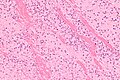
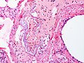
==Microscopic (VTE)== | |||
Features: | Features: | ||
* | *Layers consisting of platelets and fibrin alternating with layers of RBCs - known as ''Lines of Zahn''.<ref name=Ref_PBoD8_124>{{Ref PBoD8|124}}</ref> | ||
Note: | |||
*Multiple laminations (layers), in general, suggest that clot was formed in a dynamic environment, i.e. in the context of blood flow. | |||
===Images=== | |||
*www: | |||
**[http://library.med.utah.edu/WebPath/ATHHTML/ATH031.html Lines of Zahn (utah.edu)]. | |||
**[http://pathhsw5m54.ucsf.edu/case9/image94.html Lines of Zahn (ucsf.edu)]. | |||
<gallery> | |||
Image:Laminations_in_a_thrombus_-_low_mag.jpg | Laminated thrombus - low mag. (WC) | |||
Image:Laminations_in_a_thrombus_-_high_mag.jpg | Laminated thrombus - high mag. (WC) | |||
</gallery> | |||
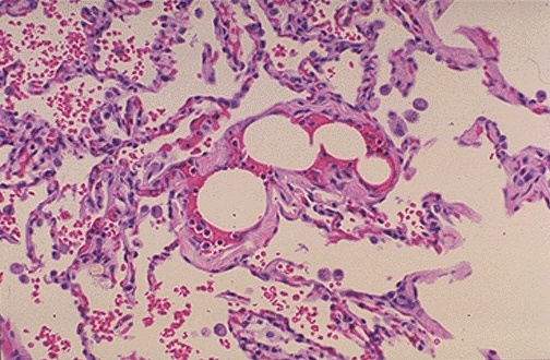
==Microscopic (fat embolism)== | |||
Features: | |||
*Fat in vessels. | |||
Images: | |||
*[http://library.med.utah.edu/WebPath/jpeg2/FOR001.jpg Fat embolism (med.utah.edu)].<ref>URL: [http://library.med.utah.edu/WebPath/EXAM/IMGQUIZ/fofrm.html http://library.med.utah.edu/WebPath/EXAM/IMGQUIZ/fofrm.html]. Accessed on: 6 December 2010.</ref> | |||
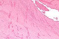
==Microscopic (amniotic fluid)== | |||
Features:<ref name=pmid13576364>{{Cite journal | last1 = ATTWOOD | first1 = HD. | title = The histological diagnosis of amniotic-fluid embolism. | journal = J Pathol Bacteriol | volume = 76 | issue = 1 | pages = 211-5 | month = Jul | year = 1958 | doi = | PMID = 13576364 }}</ref> | |||
*Pulmonary vessels with fetal debris - particularly squamous cells. | |||
Notes: | |||
*Can easily be missed.<ref name=pmid9104942>{{Cite journal | last1 = Kobayashi | first1 = H. | last2 = Ooi | first2 = H. | last3 = Hayakawa | first3 = H. | last4 = Arai | first4 = T. | last5 = Matsuda | first5 = Y. | last6 = Gotoh | first6 = K. | last7 = Tarao | first7 = T. | title = Histological diagnosis of amniotic fluid embolism by monoclonal antibody TKH-2 that recognizes NeuAc alpha 2-6GalNAc epitope. | journal = Hum Pathol | volume = 28 | issue = 4 | pages = 428-33 | month = Apr | year = 1997 | doi = | PMID = 9104942 }}</ref> | |||
*[[Alcian blue stain]] useful. (???) | |||
Images: | Images: | ||
<gallery> | |||
Image:Amniotic_fluid_embolism.jpg | Amniotic fluid embolism. (WC/Rosen) | |||
</gallery> | |||
==See also== | ==See also== | ||
*[[Pulmonary pathology]]. | *[[Pulmonary pathology]]. | ||
*[[Cholesterol embolism]]. | |||
*[[Cerebral fat embolism]]. | |||
*[[Vascular thrombus]]. | |||
*[[Intimal sarcoma]]. | |||
==References== | ==References== | ||
Latest revision as of 17:21, 18 January 2024
Pulmonary embolism, abbreviated PE, is often on the differential in autopsies, as it is not easy to diagnose clinically. Pulmonary embolism is a non-specific term; it may refer to a number of things, including:
- Pulmonary venous thromboembolism.
- Pulmonary fat embolism.
- Pulmonary foreign body embolism.
- Pulmonary septic embolism.
- Pulmonary bone marrow embolism.
- Pulmonary tumour embolism.
- Pulmonary amniotic fluid embolism.
PE usually refers to pulmonary venous thromboembolism, abbreviated VTE, if not otherwise specified.
General
- Relatively uncommon ~ 1 in 1000 adults per year.[1]
- Diagnosis in life dependent on strong clinical suspicion and radiology.
Clinical
- Shortness of breath (dyspnea) - classic symptom.
- Tachycardia.
- Chest pain.
- Findings associated with deep vein thrombosis.
- Leg pain.
- Leg swelling.
Notes:
- Venous thrombosis OR~=12 for PE.[2]
Mechanism
The classic factors are given by Virchow's triad:[3][1]
- Hypercoagulability.
- Endothelial dysfunction/injury.
- Stasis.
Note:
- The triad has a limited practical use. Like many questions about mechanism, the greatest utility, might be pimping medical students and residents.
Risks factors venous thromboembolism
A general mnemonic for hypercoagulable states PIANO:[4]
- Pregnancy.
- Immobility.
- Accidental injury.
- Nephrotic syndrome.
- Oral contraceptive pills.
Hypercoagulable states due to intrinsic causes (memory device CALM SHAPES):[5]
- Protein C deficiency.
- Antiphospholipid antibody syndrome (APLA).
- Leiden factor V deficiency.
- Malignancy.
- Protein S deficiency.
- Homocystinemia.
- Antithrombin III deficiency.
- Prothrombin G20210A.[6]
- Excess factor VIII.
- Sticky platelet syndrome.
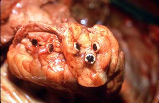
Gross (VTE)
Features:
- Intravascular spaghetti (multiple cylindrical clots - from smaller vessels) with cream sauce (gray fibrin).
- Leg swelling.
- Lines of Zahn.[7]
- Pale layers consisting of platelets and fibrin alternating with layers of RBCs; components layer during blood flow.
Notes:
- Post-mortem thrombi: one (superior) yellow portion (called "chicken fat") and one (dependent) red portion (RBCs); components layer due to gravity.
Pre- and post-mortem clots
| Feature/time | Pre-mortem | Post-mortem |
| Shininess | dull | shiny |
| Adherent to wall | yes | no |
| Colour | gray | dark purple or bilayered red/yellow |
| Pressurized | yes; "ejects itself" from lumen | no; needs to be pulled-out |
| Consistency -elastic modulus (E) -fracture toughness (K) |
firm (high E) brittle (low K) |
jello (low E) elastic (high K) |
| Image - gross | thrombus (pathguy.com), thrombus (thrombosisadviser.com) |
coronary thrombus (luc.edu)[8] |
| Image - micro. | pre- & post-mortem (elsevier.es)[9] | thrombus (oxfordjournals.org), thrombi (ucsf.edu) |
Microscopic (VTE)
Features:
- Layers consisting of platelets and fibrin alternating with layers of RBCs - known as Lines of Zahn.[7]
Note:
- Multiple laminations (layers), in general, suggest that clot was formed in a dynamic environment, i.e. in the context of blood flow.
Images
Laminated thrombus - low mag. (WC)
Laminated thrombus - high mag. (WC)


Microscopic (fat embolism)
Features:
- Fat in vessels.
Images:
Microscopic (amniotic fluid)
Features:[11]
- Pulmonary vessels with fetal debris - particularly squamous cells.
Notes:
- Can easily be missed.[12]
- Alcian blue stain useful. (???)
Images:
Amniotic fluid embolism. (WC/Rosen)

See also
- Pulmonary pathology.
- Cholesterol embolism.
- Cerebral fat embolism.
- Vascular thrombus.
- Intimal sarcoma.
References
- ↑ 1.0 1.1 Meetoo, D.. "In too deep: understanding, detecting and managing DVT.". Br J Nurs 19 (16): 1021-7. PMID 20852464.
- ↑ Reissig A, Haase U, Schulze E, Lehmann T, Kroegel C (July 2010). "[Diagnosis and therapy of pulmonary embolism prior to death]" (in German). Dtsch. Med. Wochenschr. 135 (30): 1477–83. doi:10.1055/s-0030-1262435. PMID 20648405.
- ↑ Reitsma, PH.; Versteeg, HH.; Middeldorp, S. (Mar 2012). "Mechanistic view of risk factors for venous thromboembolism.". Arterioscler Thromb Vasc Biol 32 (3): 563-8. doi:10.1161/ATVBAHA.111.242818. PMID 22345594.
- ↑ URL: http://www.usmle-forums.com/usmle-step-1-mnemonics/252-causes-hypercoagulable-states.html. Accessed on: 8 December 2011.
- ↑ Thomas RH (November 2001). "Hypercoagulability syndromes". Arch. Intern. Med. 161 (20): 2433–9. PMID 11700155. http://archinte.highwire.org/cgi/content/full/161/20/2433.
- ↑ Online 'Mendelian Inheritance in Man' (OMIM) 176930
- ↑ 7.0 7.1 Kumar, Vinay; Abbas, Abul K.; Fausto, Nelson; Aster, Jon (2009). Robbins and Cotran pathologic basis of disease (8th ed.). Elsevier Saunders. pp. 124. ISBN 978-1416031215.
- ↑ URL: http://www.meddean.luc.edu/lumen/meded/mech/cases/case1/list.htm. Accessed on 8 October 2010.
- ↑ URL: http://www.elsevier.es/cardio_eng/ctl_servlet?_f=40&ident=13142654. Accessed on: 8 October 2010.
- ↑ URL: http://library.med.utah.edu/WebPath/EXAM/IMGQUIZ/fofrm.html. Accessed on: 6 December 2010.
- ↑ ATTWOOD, HD. (Jul 1958). "The histological diagnosis of amniotic-fluid embolism.". J Pathol Bacteriol 76 (1): 211-5. PMID 13576364.
- ↑ Kobayashi, H.; Ooi, H.; Hayakawa, H.; Arai, T.; Matsuda, Y.; Gotoh, K.; Tarao, T. (Apr 1997). "Histological diagnosis of amniotic fluid embolism by monoclonal antibody TKH-2 that recognizes NeuAc alpha 2-6GalNAc epitope.". Hum Pathol 28 (4): 428-33. PMID 9104942.