Pulmonary Langerhans cell histiocytosis
| Pulmonary Langerhans cell histiocytosis | |
|---|---|
| Diagnosis in short | |
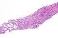
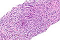
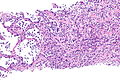
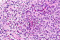
 Langerhans cell histiocytosis of the lung. H&E stain. | |
|
| |
| Synonyms | eosinophilic granuloma (of the lung) |
|
| |
| LM | cellular peribronchiolar nodules with Langerhans cells (pale staining nucleus (H&E) with nuclear infolding - "crumpled tissue paper" appearance), +/-smoker's macrophages (brown pigmented airspace macrophages), +/-eosinophilia (typical - may be rare) |
| IHC | Langerhans cells (CD1a +ve, S-100 +ve, CD207 +ve |
| Site | lung - see medical lung diseases |
|
| |
| Prevalence | uncommon |
| Radiology | upper lung zones |
| Prognosis | good with smoking cessation |
Pulmonary Langerhans cell histiocytosis is an uncommon smoking-related lung disease.
It is also known as eosinophilic granuloma of the lung.
General
- Associated with smoking.[1]
- Not associated with systemic diseases of Langerhans cells (AKA Hand-Schueller-Christian disease).
Subtypes:[1]
- Cellular form.
- Fibrotic form.
One form usually predominates.
Radiology
- Upper lung zones.
Microscopic
Features:[2]
- Cellular peribronchiolar nodules with:
- Langerhans cells - key feature:
- Pale staining nucleus (H&E) with nuclear infolding - "crumpled tissue paper" appearance.
- +/-Smoker's macrophages (brown pigmented airspace macrophages).
- +/-Eosinophilia (may be rare) - significantly narrow DDx.
- Chronic inflammatory cells (lymphocytes). (???)
- Langerhans cells - key feature:
DDx:
- Non-pulmonary Langerhans cell histiocytosis.
Images
PLCH - low mag. (WC)
PLCH - intermed. mag. (WC)
PLCH - intermed. mag. (WC)
PLCH - high mag. (WC)

PLCH - very high mag. (WC)


www:
IHC
Langerhans cells:
See also
References
- ↑ 1.0 1.1 Leslie, Kevin O.; Wick, Mark R. (2004). Practical Pulmonary Pathology: A Diagnostic Approach (1st ed.). Churchill Livingstone. pp. 234. ISBN 978-0443066313.
- ↑ 2.0 2.1 2.2 Leslie, Kevin O.; Wick, Mark R. (2004). Practical Pulmonary Pathology: A Diagnostic Approach (1st ed.). Churchill Livingstone. pp. 237. ISBN 978-0443066313.