Difference between revisions of "Pseudomembranous colitis"
Jump to navigation
Jump to search
(split-out) |
(more) |
||
| Line 1: | Line 1: | ||
{{ Infobox diagnosis | |||
| Name = {{PAGENAME}} | |||
| Image = Colonic_pseudomembranes_intermed_mag.jpg | |||
| Width = | |||
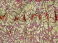
| Caption = Colonic pseudomembrane. [[H&E stain]]. | |||
| Synonyms = | |||
| Micro = heaped necrotic surface epithelium (described as "volanco lesions"), [[PMN]]s in lamina propria, +/-capillary fibrin thrombi | |||
| Subtypes = | |||
| LMDDx = [[cap polyposis]] | |||
| Stains = | |||
| IHC = | |||
| EM = | |||
| Molecular = | |||
| IF = | |||
| Gross = | |||
| Grossing = | |||
| Site = [[colon]] | |||
| Assdx = | |||
| Syndromes = | |||
| Clinicalhx = | |||
| Signs = | |||
| Symptoms = +/-diarrhea | |||
| Prevalence = uncommon | |||
| Bloodwork = | |||
| Rads = | |||
| Endoscopy = pseudomembranes (pale yellow (or white) irregular, raised mucosal lesions), interlesional mucosa often near normal grossly | |||
| Prognosis = dependent on comorbidities | |||
| Other = | |||
| ClinDDx = | |||
| Tx = dependent on underlying cause, antibiotics in ''C. difficle'' - occasionally surgical resection | |||
}} | |||
'''Pseudomembranous colitis''' an inflammation of the [[colon]] ([[colitis]]) with a characteristic endoscopic/gross appearance. It is closely associated with ''C. difficle'' infectious; however, may be seen in a number of different situations. | '''Pseudomembranous colitis''' an inflammation of the [[colon]] ([[colitis]]) with a characteristic endoscopic/gross appearance. It is closely associated with ''C. difficle'' infectious; however, may be seen in a number of different situations. | ||
Revision as of 02:05, 13 January 2014
| Pseudomembranous colitis | |
|---|---|
| Diagnosis in short | |
 Colonic pseudomembrane. H&E stain. | |
|
| |
| LM | heaped necrotic surface epithelium (described as "volanco lesions"), PMNs in lamina propria, +/-capillary fibrin thrombi |
| LM DDx | cap polyposis |
| Site | colon |
|
| |
| Symptoms | +/-diarrhea |
| Prevalence | uncommon |
| Endoscopy | pseudomembranes (pale yellow (or white) irregular, raised mucosal lesions), interlesional mucosa often near normal grossly |
| Prognosis | dependent on comorbidities |
| Treatment | dependent on underlying cause, antibiotics in C. difficle - occasionally surgical resection |
Pseudomembranous colitis an inflammation of the colon (colitis) with a characteristic endoscopic/gross appearance. It is closely associated with C. difficle infectious; however, may be seen in a number of different situations.
General
- Pseudomembranous colitis is a histomorphologic description which has a DDx. In other words, it can be caused by a number of things.
DDx of pseudomembranous colitis:[1]
- C. difficile.
- Known as C. difficile colitis.
- Ischemic colitis.
- Volvulus.
- Other infections.
Etiology:
- Anything that causes a severe mucosal injury.
Gross
Features:[2]
- Pseudomembranes:
- Pale yellow (or white) irregular, raised mucosal lesions.
- Early lesions: typical <10 mm.
- Interlesional mucosa often near normal grossly.
Images
Pseudomembranous colitis. (WC)

Microscopic
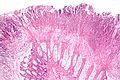
Features:[1]
- Heaped necrotic surface epithelium.
- Described as "volanco lesions" - this is what is seen endoscopically.
- PMNs in lamina propria.
- +/-Capillary fibrin thrombi.
Notes:
- Pseudomembranes arise from the crypts.
- Rarely have (benign) signet ring cell-like cells.[3]
DDx:
- Cap polyposis - very rare.
Images
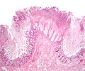
Pseudomembranes - low mag. (WC/Nephron)
Pseudomembranes - intermed. mag. (WC/Nephron)

www:
See also
References
- ↑ 1.0 1.1 Cotran, Ramzi S.; Kumar, Vinay; Fausto, Nelson; Nelso Fausto; Robbins, Stanley L.; Abbas, Abul K. (2005). Robbins and Cotran pathologic basis of disease (7th ed.). St. Louis, Mo: Elsevier Saunders. pp. 837-8. ISBN 0-7216-0187-1.
- ↑ URL: http://radiology.uchc.edu/eAtlas/GI/1749.htm. Accessed on: 22 May 2012.
- ↑ Abdulkader, I.; Cameselle-Teijeiro, J.; Forteza, J. (Apr 2003). "Signet-ring cells associated with pseudomembranous colitis.". Virchows Arch 442 (4): 412-4. doi:10.1007/s00428-003-0779-1. PMID 12684766.