Difference between revisions of "Pleomorphic adenoma"
(take out link) |
|||
| (20 intermediate revisions by the same user not shown) | |||
| Line 1: | Line 1: | ||
{{ Infobox diagnosis | {{ Infobox diagnosis | ||
| Name = {{PAGENAME}} | | Name = {{PAGENAME}} | ||
| Image = Pleomorphic_adenoma_%281%29_parotid_gland.jpg | | Image = Pleomorphic_adenoma_%281%29_parotid_gland.jpg | ||
| Width = | | Width = | ||
| Caption = Pleomorphic adenoma. [[H&E stain]]. | | Caption = Pleomorphic adenoma. [[H&E stain]]. (WC/KGH) | ||
| Synonyms = benign mixed tumour | |||
| Micro = proliferation of myoepithelium in a mesenchymal stroma +/- epithelium; cells in ducts = epithelial, cells not in ducts = myoepithelial, mesenchymal stroma = [[chondromyxoid stroma|chondroid]] stroma (specific), others (require epithelium) mucochondroid, hyalinized, osseous, fatty, myxoid | | Micro = proliferation of myoepithelium in a mesenchymal stroma +/- epithelium; cells in ducts = epithelial, cells not in ducts = myoepithelial, mesenchymal stroma = [[chondromyxoid stroma|chondroid]] stroma (specific), others (require epithelium) mucochondroid, hyalinized, osseous, fatty, myxoid | ||
| Subtypes = | | Subtypes = | ||
| LMDDx = [[myoepithelioma]], [[carcinoma ex pleomorphic adenoma]], [[epithelial-myoepithelial carcinoma]] | | LMDDx = [[myoepithelioma]], [[carcinoma ex pleomorphic adenoma]], [[epithelial-myoepithelial carcinoma]], [[polymorphous low-grade adenocarcinoma]], [[adenoid cystic carcinoma]] | ||
| Stains = | | Stains = | ||
| IHC = S-100 +ve, SMA +ve, GFAP +ve | | IHC = S-100 +ve, SMA +ve, GFAP +ve | ||
| Line 14: | Line 15: | ||
| Gross = | | Gross = | ||
| Grossing = | | Grossing = | ||
| Site = [[salivary gland]] - usu. parotid gland, other sites | | Site = [[salivary gland]] - usu. parotid gland, [[oral cavity]], other sites | ||
| Assdx = | | Assdx = | ||
| Syndromes = | | Syndromes = | ||
| Line 30: | Line 31: | ||
'''Pleomorphic adenoma''', abbreviated '''PA''', is a very common benign [[salivary gland]] tumour. | '''Pleomorphic adenoma''', abbreviated '''PA''', is a very common benign [[salivary gland]] tumour. | ||
It is also known as '''benign mixed tumour'''.<ref>URL: [http://radiopaedia.org/articles/pleomorphic-adenoma-of-the-salivary-glands http://radiopaedia.org/articles/pleomorphic-adenoma-of-the-salivary-glands]. Accessed on: 30 March 2016.</ref><ref name=pmid19926180>{{Cite journal | last1 = Lingam | first1 = RK. | last2 = Daghir | first2 = AA. | last3 = Nigar | first3 = E. | last4 = Abbas | first4 = SA. | last5 = Kumar | first5 = M. | title = Pleomorphic adenoma (benign mixed tumour) of the salivary glands: its diverse clinical, radiological, and histopathological presentation. | journal = Br J Oral Maxillofac Surg | volume = 49 | issue = 1 | pages = 14-20 | month = Jan | year = 2011 | doi = 10.1016/j.bjoms.2009.09.014 | PMID = 19926180 }}</ref> | |||
==General== | ==General== | ||
Features: | Features: | ||
| Line 44: | Line 46: | ||
==Gross== | ==Gross== | ||
*May | *May have [[cartilage|cartilaginous]] appearance. | ||
*Typically well-circumscribed. | |||
Image: | Image: | ||
| Line 70: | Line 73: | ||
*[[Carcinoma ex pleomorphic adenoma]]. | *[[Carcinoma ex pleomorphic adenoma]]. | ||
*[[Epithelial-myoepithelial carcinoma]]. | *[[Epithelial-myoepithelial carcinoma]]. | ||
*[[Polymorphous low-grade adenocarcinoma]]. | |||
*[[Adenoid cystic carcinoma]]. | |||
*[[Mucoepidermoid carcinoma]] - occasionally.<ref name=pmid15754364>{{Cite journal | last1 = Siddiqui | first1 = NH. | last2 = Wu | first2 = SJ. | title = Fine-needle aspiration biopsy of cystic pleomorphic adenoma with adnexa-like differentiation mimicking mucoepidermoid carcinoma: a case report. | journal = Diagn Cytopathol | volume = 32 | issue = 4 | pages = 229-32 | month = Apr | year = 2005 | doi = 10.1002/dc.20215 | PMID = 15754364 }}</ref> | |||
===Images=== | ===Images=== | ||
====Case 1==== | |||
<gallery> | <gallery> | ||
Image:Pleomorphic_adenoma_%281%29_parotid_gland.jpg | PA. (WC) | Image:Pleomorphic_adenoma_%281%29_parotid_gland.jpg | PA. (WC/KGH) | ||
Image:Pleomorphic_adenoma_%282%29_parotid_gland.jpg | PA. (WC) | Image:Pleomorphic_adenoma_%282%29_parotid_gland.jpg | PA. (WC/KGH) | ||
Image:Pleomorphic_adenoma_%283%29_parotid_gland.jpg | PA. (WC) | Image:Pleomorphic_adenoma_%283%29_parotid_gland.jpg | PA. (WC/KGH) | ||
Image:Pleomorphic_adenoma_%284%29_parotid_gland.jpg | PA. (WC) | Image:Pleomorphic_adenoma_%284%29_parotid_gland.jpg | PA. (WC/KGH) | ||
</gallery> | </gallery> | ||
www | ====Case 2==== | ||
<gallery> | |||
Image: Pleomorphic adenoma - cell block -- intermed mag.jpg | PA - intermed. mag. (WC) | |||
Image: Pleomorphic adenoma - cell block -- high mag.jpg | PA - intermed. mag. (WC) | |||
Image: Pleomorphic adenoma - cell block -- very high mag.jpg | PA - intermed. mag. (WC) | |||
</gallery> | |||
====www==== | |||
*[http://www.webpathology.com/image.asp?n=7&Case=111 PA - myxoid stroma (webpathology.com)]. | *[http://www.webpathology.com/image.asp?n=7&Case=111 PA - myxoid stroma (webpathology.com)]. | ||
==IHC== | ==IHC== | ||
*S-100 +ve | *S-100 +ve. | ||
*SMA +ve. | |||
*GFAP +ve. | |||
==Sign out== | ==Sign out== | ||
<pre> | |||
Left Partial Parotid, Partial Parotidectomy: | |||
- Pleomorphic adenoma. | |||
- Three benign lymph nodes. | |||
- NEGATIVE for malignancy. | |||
</pre> | |||
===Block letters=== | |||
<pre> | <pre> | ||
PAROTID GLAND MASS, RIGHT, EXCISION: | PAROTID GLAND MASS, RIGHT, EXCISION: | ||
| Line 96: | Line 119: | ||
===Micro=== | ===Micro=== | ||
The sections show a lesion with an epithelial component, on a background of a chondromyxoid stroma | The sections show a lesion with spindled (myoepithelial) cells and an epithelial component, on a background of a chondromyxoid stroma. The lesion is encapsulated by a thin layer of fibrous tissue. No nuclear atypia is apparent. Mitotic activity is not identified. | ||
Unremarkable parotid gland and lymph nodes are present. | |||
====Alternate==== | |||
The sections show a lesion with spindled (myoepithelial) cells and an epithelial component, | |||
on a background of a myxoid stroma. The lesion is mostly encapsulated by a thin layer of | |||
fibrous tissue. A small focus of macrophages is present. Significant nuclear atypia is | |||
not identified. Mitotic activity is not apparent. Unremarkable parotid gland and a | |||
morphologically benign lymph node are present. Ink is seen on the tumour. | |||
====Biopsy==== | |||
The sections show a lesion with spindled (myoepithelial) cells and an epithelial component, on a background of a chondromyxoid stroma. No nuclear atypia is apparent. Mitotic activity is not identified. | |||
<pre> | |||
The tumour stains as follows: | |||
POSITIVE: CK7, S-100, SMA, GFAP (patchy). | |||
NEGATIVE: (none). | |||
PROLIFERATION (Ki-67): <2%. | |||
</pre> | |||
==See also== | ==See also== | ||
*[[Salivary glands]]. | *[[Salivary glands]]. | ||
*[[Salivary gland cytopathology]]. | |||
==References== | ==References== | ||
Latest revision as of 16:21, 30 November 2023
| Pleomorphic adenoma | |
|---|---|
| Diagnosis in short | |
_parotid_gland.jpg) Pleomorphic adenoma. H&E stain. (WC/KGH) | |
|
| |
| Synonyms | benign mixed tumour |
|
| |
| LM | proliferation of myoepithelium in a mesenchymal stroma +/- epithelium; cells in ducts = epithelial, cells not in ducts = myoepithelial, mesenchymal stroma = chondroid stroma (specific), others (require epithelium) mucochondroid, hyalinized, osseous, fatty, myxoid |
| LM DDx | myoepithelioma, carcinoma ex pleomorphic adenoma, epithelial-myoepithelial carcinoma, polymorphous low-grade adenocarcinoma, adenoid cystic carcinoma |
| IHC | S-100 +ve, SMA +ve, GFAP +ve |
| Site | salivary gland - usu. parotid gland, oral cavity, other sites |
|
| |
| Signs | mass lesion |
| Prevalence | common |
| Prognosis | benign |
| Clin. DDx | other salivary gland tumours |
Pleomorphic adenoma, abbreviated PA, is a very common benign salivary gland tumour.
It is also known as benign mixed tumour.[1][2]
General
Features:
- Very common - approx. 60% of parotid gland tumours.[3]
- May transform into a malignant tumour.
- Other benign salivary gland tumours do not do this.
- Only benign childhood salivary gland tumour of significance.
Weinreb's dictums
- Most common salivary tumour in all age groups.
- Seen in all sites (unlike other benign tumours).
- Recurrence and malignancy risk (unlike other benign salivary gland tumours).
- Any part of a tumour that looks like PA makes it a PA.
Gross
- May have cartilaginous appearance.
- Typically well-circumscribed.
Image:
Microscopic
Features:[3]
- Proliferation of myoepithelium and epithelium (ductal cells) in mesenchymal stroma.
- Cells in ducts = epithelial.
- Cells not in ducts = myoepithelial.[4]
- Mesenchymal stroma - important feature.
Notes:
- Mesenchymal stroma not required for diagnosis -- if >5% ducts.[4]
- No chondroid stroma and <5% ductal cells = myoepithelioma.
- Look for, i.e. rule-out, poorly differentiated carcinoma: carcinoma ex pleomorphic adenoma.
Memory device: MEC = myoepithelium, epithelium, chondromyxoid stroma.
DDx:
- Myoepithelioma.
- Carcinoma ex pleomorphic adenoma.
- Epithelial-myoepithelial carcinoma.
- Polymorphous low-grade adenocarcinoma.
- Adenoid cystic carcinoma.
- Mucoepidermoid carcinoma - occasionally.[5]
Images
Case 1
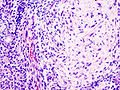
PA. (WC/KGH)
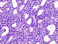
PA. (WC/KGH)
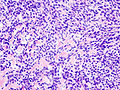
PA. (WC/KGH)
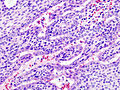
PA. (WC/KGH)
_parotid_gland.jpg)
_parotid_gland.jpg)
_parotid_gland.jpg)
Case 2
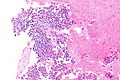
PA - intermed. mag. (WC)
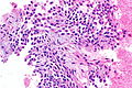
PA - intermed. mag. (WC)
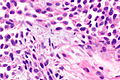
PA - intermed. mag. (WC)


www
IHC
- S-100 +ve.
- SMA +ve.
- GFAP +ve.
Sign out
Left Partial Parotid, Partial Parotidectomy:
- Pleomorphic adenoma.
- Three benign lymph nodes.
- NEGATIVE for malignancy.
Block letters
PAROTID GLAND MASS, RIGHT, EXCISION:
- PLEOMORPHIC ADENOMA.
- FOUR BENIGN LYMPH NODES.
- NEGATIVE FOR MALIGNANCY.
Note:
- Complete excision is often elusive; stating "completely excised" on a surgical pathology report is unwise.[4]
Micro
The sections show a lesion with spindled (myoepithelial) cells and an epithelial component, on a background of a chondromyxoid stroma. The lesion is encapsulated by a thin layer of fibrous tissue. No nuclear atypia is apparent. Mitotic activity is not identified.
Unremarkable parotid gland and lymph nodes are present.
Alternate
The sections show a lesion with spindled (myoepithelial) cells and an epithelial component, on a background of a myxoid stroma. The lesion is mostly encapsulated by a thin layer of fibrous tissue. A small focus of macrophages is present. Significant nuclear atypia is not identified. Mitotic activity is not apparent. Unremarkable parotid gland and a morphologically benign lymph node are present. Ink is seen on the tumour.
Biopsy
The sections show a lesion with spindled (myoepithelial) cells and an epithelial component, on a background of a chondromyxoid stroma. No nuclear atypia is apparent. Mitotic activity is not identified.
The tumour stains as follows:
POSITIVE: CK7, S-100, SMA, GFAP (patchy).
NEGATIVE: (none).
PROLIFERATION (Ki-67): <2%.
See also
References
- ↑ URL: http://radiopaedia.org/articles/pleomorphic-adenoma-of-the-salivary-glands. Accessed on: 30 March 2016.
- ↑ Lingam, RK.; Daghir, AA.; Nigar, E.; Abbas, SA.; Kumar, M. (Jan 2011). "Pleomorphic adenoma (benign mixed tumour) of the salivary glands: its diverse clinical, radiological, and histopathological presentation.". Br J Oral Maxillofac Surg 49 (1): 14-20. doi:10.1016/j.bjoms.2009.09.014. PMID 19926180.
- ↑ 3.0 3.1 Thompson, Lester D. R. (2006). Head and Neck Pathology: A Volume in Foundations in Diagnostic Pathology Series (1st ed.). Churchill Livingstone. pp. 295. ISBN 978-0443069604.
- ↑ 4.0 4.1 4.2 Weinreb I. 10 January 2011.
- ↑ Siddiqui, NH.; Wu, SJ. (Apr 2005). "Fine-needle aspiration biopsy of cystic pleomorphic adenoma with adnexa-like differentiation mimicking mucoepidermoid carcinoma: a case report.". Diagn Cytopathol 32 (4): 229-32. doi:10.1002/dc.20215. PMID 15754364.