Difference between revisions of "Pituitary gland"
Jump to navigation
Jump to search
(→Rathke cleft cyst: mv from H&N article) |
m (→General: tweak) |
||
| Line 80: | Line 80: | ||
===General=== | ===General=== | ||
*Benign counterpart of craniopharyngioma | *Benign counterpart of craniopharyngioma | ||
*Arises from [[pituitary gland]] | *Arises from intermediate lobe of [[pituitary gland]] (''pars intermedia of pituitary gland''). | ||
Radiology: | Radiology: | ||
Revision as of 15:49, 5 December 2010
The pituitary gland is known as the master gland.
Divisions:[1]
- Anterior pituitary (AKA adenohypophysis).
- Posterior pituitary (AKA neurohypophysis, neural pituitary).
Function
Anterior
Hormones:[2]
- Growth hormone (GH).
- Luteinizing hormone (LH)
- Follicle-stimulating hormone (FSH)
- Thyroid stimulating hormone (TSH)
- Adrenocorticotropic hormone (ACTH)
- Prolactin (PRL)
Mnemonic: "Go Look For The Adenoma Please" = GH, LH, FSH, TSH, ACTH, PRL.
Posterior
Hormones:[2]
- Oxytocin.
- Antidiuretic hormone (ADH).
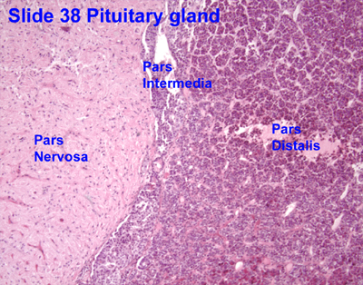
Anatomy and histology
Anatomy
Basic anatomy (simplified):[3]
- Anterior:
- Pars distalis.
- Pars intermedia.
- Posterior:
- Pars nervosa.
Embryological origin:[3]
- Anterior - Rathke's pouch (roof of mouth).
- Posterior - diencephalon (ventral aspect).
Images:
Histology
Anterior
- Acidophils (40% of cells) = red or orange.
- GH, PRL.
- Basophils (10% of cells) = basophilic (light blue).
- TSH, LH, FSH.
- Chromophobes (50% of cells) = amphophilic (purplish/grey).
Notes:
- The cellular product (i.e. hormone produced) is not strictly correlated with the cell type.[4]
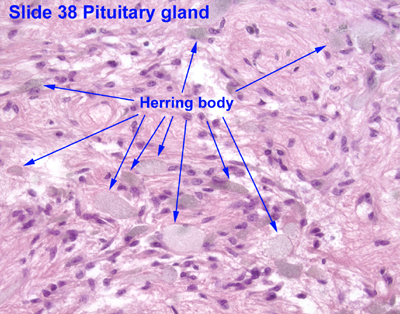
Posterior
Features:[4]
- Herring bodies - key feature.
- Eosinophilic axonal dilations filled with lysosomes and neurosecretory granules.
- Less cellular.
- Usually more cellular in perivascular location.
Image: Herring bodies (ouhsc.edu).
DDx for stellar lesions
- Pituitary adenoma.
- Rathke cleft cyst.
- Craniopharyngioma.
- Germ cell tumour.
- Meningioma.
Pituitary adenoma
General
- Classically presents with visual field defects.
Microscopic
Features:[5]
- Loss of fibrous stroma.
Notes:
- Smears very well.[6]
Rathke cleft cyst
General
- Benign counterpart of craniopharyngioma
- Arises from intermediate lobe of pituitary gland (pars intermedia of pituitary gland).
Radiology:
- Typically no calcifications.[7]
Radiologic DDx:[7]
- Arachnoid cyst.
- Craniopharyngioma.
- Cysticercosis (see microorganisms).
- Pituitary adenoma.
- Epidermoid of brain.
Microscopic
Features:
- Lined by cuboidal or columnar epithelial +occasional goblet cells.[8]
- +/-Squamous metaplasia.
Image: Rathke's cleft cyst (endotext.org).
Craniopharyngioma
- Related to Rathke cleft cyst.
Necrosis
- Rare.
Causes
- Sheehan syndrome - secondary to blood loss in childbirth.[9]
- Syphilis (fetal-maternal transmission).[10]
- Mollaret's meningitis - very rare.[11] (???)
- Spontaneous necrosis of pituitary tumours - case reports.[12]
Autoimmune hypophysitis
General
Features:[13]
- Rare.
- Autoantigens are unknown.
- May be misdiagnosed as a nonsecreting adenoma.
Microscopic
Features:[13]
- Lymphocytic infiltration.
See also
References
- ↑ http://www.vivo.colostate.edu/hbooks/pathphys/endocrine/hypopit/histo.html
- ↑ 2.0 2.1 http://users.rcn.com/jkimball.ma.ultranet/BiologyPages/P/Pituitary.html
- ↑ 3.0 3.1 URL: http://www.vivo.colostate.edu/hbooks/pathphys/endocrine/hypopit/histo_pit.html. Accessed on: 31 October 2010.
- ↑ 4.0 4.1 Perry, Arie; Brat, Daniel J. (2010). Practical Surgical Neuropathology: A Diagnostic Approach: A Volume in the Pattern Recognition series (1st ed.). Churchill Livingstone. pp. 26. ISBN 978-0443069826.
- ↑ Perry, Arie; Brat, Daniel J. (2010). Practical Surgical Neuropathology: A Diagnostic Approach: A Volume in the Pattern Recognition series (1st ed.). Churchill Livingstone. pp. 36. ISBN 978-0443069826.
- ↑ MUN. 24 November 2010.
- ↑ 7.0 7.1 URL: http://emedicine.medscape.com/article/343629-overview. Accessed on: 14 November 2010.
- ↑ URL: http://www.endotext.org/neuroendo/neuroendo3/neuroendo3.html. Accessed on: 27 May 2010.
- ↑ URL: http://www.mayoclinic.com/health/sheehans-syndrome/DS00889. Accessed on: 16 November 2010.
- ↑ URL: http://pediatrics.aappublications.org/cgi/content/full/104/1/e4. Accessed on: 16 November 2010.
- ↑ Dancer CM, Woods ML, Henderson RD, Robertson T, Mungomery M, Allworth A (July 2008). "Mollaret's meningitis and pituitary failure associated with a Rathke's cleft cyst". Intern Med J 38 (7): 609–11. doi:10.1111/j.1445-5994.2008.01709.x. PMID 18715308.
- ↑ Sachdev Y, Evered DC, Hall R (April 1976). "Spontaneous pituitary necrosis". Br Med J 1 (6015): 942. PMC 1639254. PMID 1268492. http://www.ncbi.nlm.nih.gov/pmc/articles/PMC1639254/pdf/brmedj00512-0028a.pdf.
- ↑ 13.0 13.1 Tzou SC, Lupi I, Landek M, et al. (July 2008). "Autoimmune hypophysitis of SJL mice: clinical insights from a new animal model". Endocrinology 149 (7): 3461–9. doi:10.1210/en.2007-1692. PMC 2453094. PMID 18388197. https://www.ncbi.nlm.nih.gov/pmc/articles/PMC2453094/.
External links
- Neuropathology - neuropathologyweb.org.
- Endocrine histology (anhb.uwa.edu.au).