Difference between revisions of "Paraganglioma"
Jump to navigation
Jump to search
m (→Microscopic: more) |
(+gross) |
||
| Line 8: | Line 8: | ||
*Carotid body tumour = paraganglioma of carotid body. | *Carotid body tumour = paraganglioma of carotid body. | ||
==Epidemiology== | ===Epidemiology=== | ||
*Very rare | *Very rare | ||
*Rarely malignant | *Rarely malignant | ||
| Line 18: | Line 18: | ||
**[[MEN 2B]]. | **[[MEN 2B]]. | ||
==Clinical== | ===Clinical=== | ||
*10% bilateral, multiple, familial, pediatric and malignant.<ref name=Ref_EP327>{{Ref EP|327}}</ref> | *10% bilateral, multiple, familial, pediatric and malignant.<ref name=Ref_EP327>{{Ref EP|327}}</ref> | ||
==Gross== | |||
*Dusky colour. | |||
Note: | |||
*''Pheo'' (in [[pheochromocytoma]]) is ''dusky''; ''chromo'' is ''colour''. | |||
Image: | |||
*[http://commons.wikimedia.org/wiki/File:Mediastinal_paraganglioma.jpg Mediastinal paraganglioma (WC/AFIP)]. | |||
==Microscopic== | ==Microscopic== | ||
Revision as of 04:14, 19 May 2012
Paraganglioma is a rare tumour arising from the paraganglion. A paraganglioma arising in the adrenal gland is known as a pheochromocytoma.
General
- Definition: tumour of paraganglion.
- Can be sympathetic or parasympathetic.
- Most common paraganglioma = pheochromocytoma.[1]
- Head & neck most common site - after abdomen.
- Carotid body tumour = paraganglioma of carotid body.
Epidemiology
- Very rare
- Rarely malignant
- Familial syndromes assoc. with paragangliomas.[2]
- von Hippel Lindau.
- Hereditary paragangliomatosis.
- Neurofibromatosis type 1 (von Recklinghausen disease).
- MEN 2A.
- MEN 2B.
Clinical
- 10% bilateral, multiple, familial, pediatric and malignant.[3]
Gross
- Dusky colour.
Note:
- Pheo (in pheochromocytoma) is dusky; chromo is colour.
Image:
Microscopic
Features:[4]
- Zellballen (literally: "cell balls") - nests of cells.
- Fibrovascular septae.
- Finely granular cytoplasm (salt-and-pepper nuclei).
- +/-Hemorrhage - very common.
DDx:
- Neuroendocrine tumour - nests surrounded by stroma/do not touch.
- Pheochromocytoma - paraganglioma of the adrenal gland.
Images:
- WC:
- www:
IHC
Features:[5]
- Chromogranin +ve.
- Synaptophysin +ve.
- S100 +/-.
- Cytokeratin -ve.
- EMA -ve.
- +ve in RCC.
EM
Features:[6]
- Neurosecretory granules.
- Electron dense core.
- Typically perinuclear location.
Image:
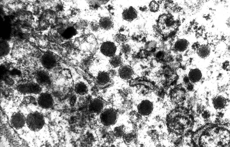
See also
References
- ↑ Thompson, Lester D. R. (2006). Endocrine Pathology: A Volume in Foundations in Diagnostic Pathology Series (1st ed.). Churchill Livingstone. pp. 327. ISBN 978-0443066856.
- ↑ Thompson, Lester D. R. (2006). Endocrine Pathology: A Volume in Foundations in Diagnostic Pathology Series (1st ed.). Churchill Livingstone. pp. 328. ISBN 978-0443066856.
- ↑ Thompson, Lester D. R. (2006). Endocrine Pathology: A Volume in Foundations in Diagnostic Pathology Series (1st ed.). Churchill Livingstone. pp. 327. ISBN 978-0443066856.
- ↑ Thompson, Lester D. R. (2006). Endocrine Pathology: A Volume in Foundations in Diagnostic Pathology Series (1st ed.). Churchill Livingstone. pp. 329-332. ISBN 978-0443066856.
- ↑ Thompson, Lester D. R. (2006). Endocrine Pathology: A Volume in Foundations in Diagnostic Pathology Series (1st ed.). Churchill Livingstone. pp. 335. ISBN 978-0443066856.
- ↑ 6.0 6.1 URL: http://path.upmc.edu/cases/case408.html. Accessed on: 16 January 2012.