Difference between revisions of "Papanicolaou stain"
Jump to navigation
Jump to search
(→Use) |
(→Images) |
||
| (3 intermediate revisions by the same user not shown) | |||
| Line 15: | Line 15: | ||
| Other = | | Other = | ||
}} | }} | ||
'''Papanicolaou stain''', abbreviated '''Pap stain''', is a standard [[stain]] used in [[cytopathology]].<ref>{{Cite journal | last1 = Prabhudesai | first1 = NM. | last2 = Kulkarni | first2 = MB. | last3 = Desai | first3 = SB. | last4 = Borges | first4 = AM. | title = Modified H & E staining technique for fine needle aspiration cytology (FNAC) smears. | journal = Indian J Pathol Microbiol | volume = 47 | issue = 3 | pages = 384-6 | month = Jul | year = 2004 | doi = | PMID = 16295430 }}</ref> It is a modified [[H&E stain]]. | '''Papanicolaou stain''', abbreviated '''Pap stain''', is a standard [[stain]] used in [[cytopathology]].<ref name=pmid16295430>{{Cite journal | last1 = Prabhudesai | first1 = NM. | last2 = Kulkarni | first2 = MB. | last3 = Desai | first3 = SB. | last4 = Borges | first4 = AM. | title = Modified H & E staining technique for fine needle aspiration cytology (FNAC) smears. | journal = Indian J Pathol Microbiol | volume = 47 | issue = 3 | pages = 384-6 | month = Jul | year = 2004 | doi = | PMID = 16295430 }}</ref> It is a modified [[H&E stain]]. | ||
==General== | ==General== | ||
*Can be thought of as the [[H&E stain|H&E]] of [[cytopathology]]. | *Can be thought of as the [[H&E stain|H&E]] of [[cytopathology]]. | ||
**It is a modified [[H&E stain]]. | **It is a modified [[H&E stain]]. | ||
*Specimens are fixed in ethanol. | *Specimens are [[fixation|fixed]] in ethanol. | ||
*Good for seeing nuclear detail. | *Good for seeing nuclear detail.<ref name=pmid16295430/> | ||
*Out-of-focus cytoplasm is translucent; allows one to focus overlapped cells in different planes. | *Out-of-focus cytoplasm is translucent; allows one to focus overlapped cells in different planes. | ||
| Line 34: | Line 34: | ||
===Images=== | ===Images=== | ||
<gallery> | <gallery> | ||
Image:Low grade squamous intraepithelial lesion.jpg|LSIL - Pap stain. (WC) | Image:Low grade squamous intraepithelial lesion.jpg|Cervix - LSIL - Pap stain. (WC) | ||
Image:High-grade squamous intraepithelial lesion.jpg|HSIL - Pap stain. (WC) | Image:High-grade squamous intraepithelial lesion.jpg|Cervix - HSIL - Pap stain. (WC) | ||
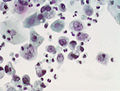
Image:Lung small cell carcinoma -- extremely high mag.jpg|Lung - SmCC - Pap stain. (WC) | |||
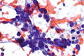
Image:Urine_citology_urothelial_carcinoma_2.jpg | [[Urine cytology]] - UCC - Pap stain. (WC) | |||
Image:Urine_citology_urothelial_carcinoma_2.jpg | [[Urine cytology]] - Pap stain. (WC) | |||
</gallery> | </gallery> | ||
Latest revision as of 04:17, 24 April 2016
| Papanicolaou stain | |
|---|---|
| Stain in short | |
 Low grade squamous intraepithelial lesion. | |
| Abbreviation | Pap stain |
| Similar stains | Romanowsky stains |
| Use | the standard stain in cytopathology |
| Interpretation | blue/purple = nucleus, pink/green = cytoplasm, orange = keratin |
Papanicolaou stain, abbreviated Pap stain, is a standard stain used in cytopathology.[1] It is a modified H&E stain.
General
- Can be thought of as the H&E of cytopathology.
- It is a modified H&E stain.
- Specimens are fixed in ethanol.
- Good for seeing nuclear detail.[1]
- Out-of-focus cytoplasm is translucent; allows one to focus overlapped cells in different planes.
Use
Interpretation
- Blue/purple = nucleus.
- Green/pink = cytoplasm.
- Orange = keratin.
Images
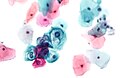
Cervix - LSIL - Pap stain. (WC)
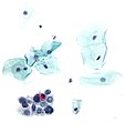
Cervix - HSIL - Pap stain. (WC)
Lung - SmCC - Pap stain. (WC)
Urine cytology - UCC - Pap stain. (WC)


