Difference between revisions of "Panniculitis"
Jump to navigation
Jump to search
m (→Microscopic) |
|||
| Line 52: | Line 52: | ||
Notes: | Notes: | ||
#No [[vasculitis]]. | #No [[vasculitis]]. | ||
#+/-Granulomas.<ref name=uscf_en>URL: [http://missinglink.ucsf.edu/lm/DermatologyGlossary/erythema_nodosum.html http://missinglink.ucsf.edu/lm/DermatologyGlossary/erythema_nodosum.html]. Accessed on: 11 September 2011.</ref> | #+/-Granulomas - within the septa.<ref name=uscf_en>URL: [http://missinglink.ucsf.edu/lm/DermatologyGlossary/erythema_nodosum.html http://missinglink.ucsf.edu/lm/DermatologyGlossary/erythema_nodosum.html]. Accessed on: 11 September 2011.</ref> | ||
DDx: | DDx: | ||
| Line 60: | Line 60: | ||
*[http://missinglink.ucsf.edu/lm/DermatologyGlossary/img/Dermatology%20Glossary/Glossary%20Histo%20Images/Erythema_Nodosum_low_power.jpg EN - low mag. (ucsf.edu)].<ref name=uscf_en>URL: [http://missinglink.ucsf.edu/lm/DermatologyGlossary/erythema_nodosum.html http://missinglink.ucsf.edu/lm/DermatologyGlossary/erythema_nodosum.html]. Accessed on: 11 September 2011.</ref> | *[http://missinglink.ucsf.edu/lm/DermatologyGlossary/img/Dermatology%20Glossary/Glossary%20Histo%20Images/Erythema_Nodosum_low_power.jpg EN - low mag. (ucsf.edu)].<ref name=uscf_en>URL: [http://missinglink.ucsf.edu/lm/DermatologyGlossary/erythema_nodosum.html http://missinglink.ucsf.edu/lm/DermatologyGlossary/erythema_nodosum.html]. Accessed on: 11 September 2011.</ref> | ||
*[http://missinglink.ucsf.edu/lm/DermatologyGlossary/img/Dermatology%20Glossary/Glossary%20Histo%20Images/Erythema_Nodosum_high_power.jpg EN - high mag. (ucsf.edu)]. | *[http://missinglink.ucsf.edu/lm/DermatologyGlossary/img/Dermatology%20Glossary/Glossary%20Histo%20Images/Erythema_Nodosum_high_power.jpg EN - high mag. (ucsf.edu)]. | ||
*[http://dermaamin.com/site/histopathology-of-the-skin/57-e/1720-erythema-nodosum-.html EN - several images (dermaamin.com)]. | |||
==Morphea profunda== | ==Morphea profunda== | ||
Revision as of 17:16, 17 October 2012
Panniculitis is inflammation of the adipose tissue and fits into the larger category of inflammatory skin disorders. It is encountered in dermatopathology specimens.
Classification
- Lobular - involve fat lobules.
- Septal - involve interlobular septae.
A simple general DDx
Septal:
Lobular:
- Erythema induratum.
- Self-inflicted trauma (factitial panniculitis).
- Systemic lupus erythematosus.
- Other.
DDx by type
Septal:
- Erythema nodosum.
- Scleroderma panniculitis, also morphea profunda.[1]
Lobular:[1]
- Infectious panniculitis.
- Erythema induratum.
- Lupus panniculitis.
- Pancreatic panniculitis.
- Alpha1-antitrypsin deficiency.
- Subcutaneous fat necrosis of the newborn.
- Sclerema neonatorum.
Specific conditions
Erythema nodosum
Causes - mnemonic NODOSUM:[2]
- NO cause (idiopathic) ~ 55% of cases.
- Drugs (sulfonamides, amoxicillin, oral contraceptives) ~ 5% of cases.
- Other infections - group A streptococci (streptococcal pharyngitis), Yersinia, chlamydia, mycobacteria, others ~ 30% of cases.
- Sarcoidosis ~ 7% of cases.
- Ulcerative colitis & Crohn's disease ~ 2% of cases.
- Malignancy (leukemia, Hodgkin's lymphoma) ~ 1% of cases
Microscopic
- Expanded septa between fat lobules - key (low power) feature.
- Neutrophils.
- Lymphocytes.
- Histiocytes.
- Fibrin.
Notes:
- No vasculitis.
- +/-Granulomas - within the septa.[6]
DDx:
Images:
Morphea profunda
- AKA scleroderma panniculitis.
General
Main article: Scleroderma
- Think about the name... sclerotic (sclero-) dermis (-derma).
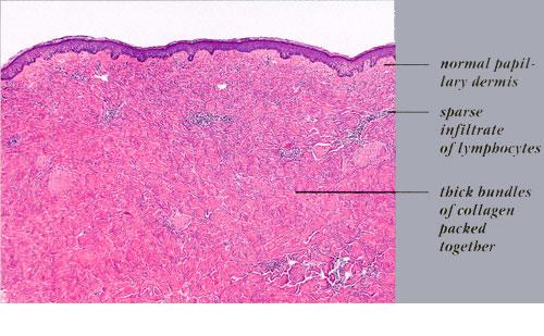
Microscopic
Features:[1]
- Septal fibrosis/expansion - key feature.
- Classically ~2-3x an adipocytes thick.
- +/-Deep dermal perivascular lymphocytes and plasma cells.
DDx:
- Erythema nodosum.
- Lichen sclerosus - fibrosis only superficial.
Images:
Erythema induratum
General
Features:[4]
- Uncommon.
- Etiology: unknown.
- Brinster[1] suggests an association, in some cases, with Mycobacterium tuberculosis and hepatitis C.
Clinical:[5]
- Classic location: posterior shins.
- Ulcerates and scars.
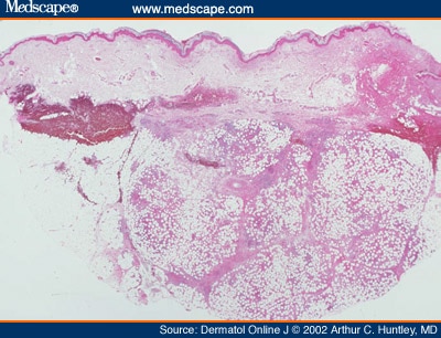
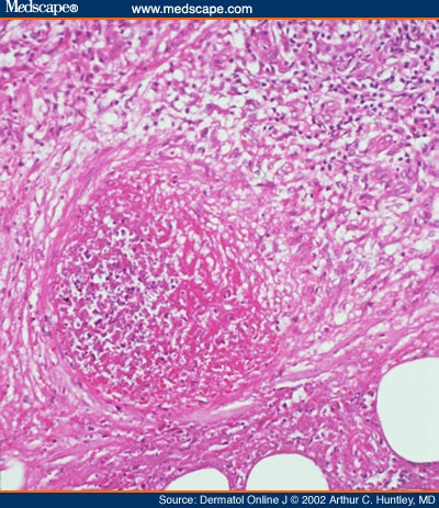
Microscopic
Features:[4]
- Predominantly lobular process with:[3]
- Necrotizing granulomatous inflammation.
- Necrotizing vasculitis of small/medium sized vessels (early).
DDx:
- Granulomatous infection.
- Vasculitis.
Images:
Stains
- AFB stain - exclude Tuberculosis.
See also
References
- ↑ 1.0 1.1 1.2 1.3 Brinster, NK. (Nov 2008). "Dermatopathology for the surgical pathologist: a pattern-based approach to the diagnosis of inflammatory skin disorders (part II).". Adv Anat Pathol 15 (6): 350-69. doi:10.1097/PAP.0b013e31818b1ac6. PMID 18948765.
- ↑ 2.0 2.1 Schwartz, RA.; Nervi, SJ. (Mar 2007). "Erythema nodosum: a sign of systemic disease.". Am Fam Physician 75 (5): 695-700. PMID 17375516.
- ↑ 3.0 3.1 URL: http://www.medscape.com/viewarticle/440356_8. Accessed on: 11 September 2011.
- ↑ 4.0 4.1 4.2 Kumar, Vinay; Abbas, Abul K.; Fausto, Nelson; Aster, Jon (2009). Robbins and Cotran pathologic basis of disease (8th ed.). Elsevier Saunders. pp. 1199. ISBN 978-1416031215.
- ↑ 5.0 5.1 Mitchell, Richard; Kumar, Vinay; Fausto, Nelson; Abbas, Abul K.; Aster, Jon (2011). Pocket Companion to Robbins & Cotran Pathologic Basis of Disease (8th ed.). Elsevier Saunders. pp. 609. ISBN 978-1416054542.
- ↑ 6.0 6.1 URL: http://missinglink.ucsf.edu/lm/DermatologyGlossary/erythema_nodosum.html. Accessed on: 11 September 2011.
- ↑ URL: http://www.dermaamin.com/site/histopathology-of-the-skin/71-s/2154-superficial-morphea-.html. Accessed on: 5 October 2011.