Paget's disease of the breast
Paget's disease of the breast, also Paget disease of the breast and Paget's disease of the nipple, is a lesion of the breast. It is abbreviated PDB.[1]
| Paget's disease of the breast | |
|---|---|
| Diagnosis in short | |
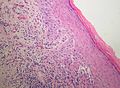
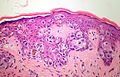
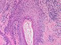
 Paget's disease. H&E stain. | |
|
| |
| LM | large epithelioid cells - nested or single - in the epidermis, clear/pale cytoplasm (occasionally eosinophilic), large nucleoli |
| LM DDx | benign Toker cell hyperplasia, malignant melanoma, Bowen's disease, apocrine carcinoma of the skin |
| IHC | CK7 +ve, CEA +ve, S-100 -ve, CK5/6 -ve, HER2 +ve |
| Gross | erythema, +/-weeping, +/-crusted |
| Site | breast |
|
| |
| Prognosis | usu. associated with invasive breast carcinoma |
| Clin. DDx | nipple adenoma, contact dermatitis |
There is also a Paget's disease of the bone - just to make things confusing. This is dealt with in its own article and has nothing (from a pathologic perspective) to do with the Paget's disease discussed in this article; these two things just happened to be discovered by the same guy.
Non-bone Paget's disease is subdivided into:
- Mammary Paget's disease - dealt with in this article.
- Extramammary Paget's disease.
Histologically, i.e. under the microscope, the above are essentially identically; however, the associations (and prognosis) are quite different!
General
- Cells in the epithelium, i.e. skin, that look like they don't belong.
- Associated with underlying invasive breast carcinoma.[2]
Note:
- Extramammary Paget's disease is not usually associated with malignancy.
Gross
Features:[3]
- Erythematous, i.e. red.
- +/-Crusted.
- +/-Weeping (wet).
DDx - clinical:
Images:
Microscopic
Features:[2]
- Epitheliod morphology (round/ovoid).
- Cells nested or single.
- Clear/pale cytoplasm key feature - may also be eosinophilic.
- Large nucleoli.
Note:
- The neoplastic cells of Paget's disease may contain melanin.[7]
DDx
- Benign Toker cell hyperplasia.
- Malignant melanoma.
- Bowen disease.
- Nipple (duct) adenoma (clinical DDx).
- Apocrine carcinoma of the skin.[8]
Images
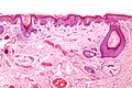
Paget disease - low mag. (WC/Nephron)
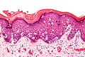
Paget disease - high mag. (WC/Nephron)
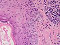
Breast Paget Disease - high power - (SKB)
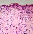
Breast Paget Disease - underlying invasive carcinoma - medium power - (SKB)
Breast Paget Disease - high power - (SKB)
Breast Paget Disease - high power - (SKB)
Breast Paget Disease - medium power - (SKB)


www:
IHC
Panel:[2]
- S-100 -ve, HMB-45 -ve (both typically +ve in melanoma).
- CK7 +ve. (???)
- Toker cells CK7 +ve.[9]
- CEA +ve (-ve in Bowen's disease, -ve in Toker cells).
Additional:
Tabular comparison
IHC features of Paget disease and its DDx:[12][13]
| Entity | CK5/6 | CK7 | CAM5.2 | EMA | CEA | HER2 | S100 | HMB-45 |
|---|---|---|---|---|---|---|---|---|
| Paget disease | CK5/6 -ve | CK7 +ve (?) | CAM5.2 +ve | EMA +ve | CEA +ve | HER2 +ve | S100 -ve | HMB-45 -ve |
| Bowen disease | CK5/6 +ve | CK7 -ve | CAM5.2 -ve | EMA -ve | CEA -ve | HER2 -ve (?) | S100 -ve | HMB-45 -ve |
| Toker cell hyperplasia | CK5/6 ? | CK7 +ve | CAM5.2 ? | EMA +ve/-ve[14] | CEA -ve[15] | HER2 -ve[9] | S100 -ve[15] | HMB-45 ? |
| Melanoma | CK5/6 -ve | CK7 -ve | CAM5.2 -ve | EMA -ve | CEA -ve | HER2 -ve | S100 +ve | HMB-45 +ve |
Mini-table comparison
| Entity | CK7 | CEA | HER2 |
|---|---|---|---|
| Paget disease | CK7 +ve | CEA +ve | HER2 +ve |
| Toker cell hyperplasia | CK7 +ve | CEA -ve | HER2 -ve |
| Bowen disease | CK7 -ve | CEA -ve | HER2 -ve |
Sign out
Right Nipple, Biopsy:
- Paget's disease.

Comment:
The Paget-like cells stain as follows:
POSITIVE: CK7 (moderate, diffuse), HER2 (complete membranous, moderate/strong).
NEGATIVE: S-100, HMB-45, CK5, CEA-mono.

The morphologic and immunohistochemical findings are in keeping with Paget's disease of the breast.

The breast imaging findings are noted.
See also
References
- ↑ Ellis, PE.; Maclean, AB.; Crow, JC.; Wong Te Fong, LF.; Rolfe, KJ.; Perrett, CW. (Dec 2009). "Expression of cyclin D1 and retinoblastoma protein in Paget's disease of the vulva and breast: an immunohistochemical study of 108 cases.". Histopathology 55 (6): 709-15. doi:10.1111/j.1365-2559.2009.03434.x. PMID 19919588.
- ↑ 2.0 2.1 2.2 URL: http://emedicine.medscape.com/article/1101235-diagnosis
- ↑ Lefkowitch, Jay H. (2006). Anatomic Pathology Board Review (1st ed.). Saunders. pp. 294 & 306 Q15. ISBN 978-1416025887.
- ↑ URL: http://www.danderm-pdv.is.kkh.dk/atlas/2-38.html. Accessed on: 30 May 2012.
- ↑ Feroze, K.; Manoj, J.; Venkitakrishnan, S. (2008). "Allergic contact dermatitis mimicking mammary paget's disease.". Indian J Dermatol 53 (3): 154-5. doi:10.4103/0019-5154.43210. PMID 19882019.
- ↑ URL: http://www.gfmer.ch/selected_images_v2/search_result_list.php?offset=15¶m1=Paget%20disease%20of%20breast¶m2=and. Accessed on: 30 May 2012.
- ↑ Lefkowitch, Jay H. (2006). Anatomic Pathology Board Review (1st ed.). Saunders. pp. 306-7 Q15. ISBN 978-1416025887.
- ↑ URL: http://derm101.com/searchResults.aspx?searchStr=apocrine+carcinoma&rootTerm=apocrine+carcinoma&searchType=2&rootID=12687. Accessed on: 9 September 2011.
- ↑ 9.0 9.1 Nofech-Mozes, S.; Hanna, W.. "Toker cells revisited.". Breast J 15 (4): 394-8. doi:10.1111/j.1524-4741.2009.00743.x. PMID 19601945.
- ↑ RS. May 2010.
- ↑ Khayyata, S.; Yun, S.; Pasha, T.; Jian, B.; McGrath, C.; Yu, G.; Gupta, P.; Baloch, Z. (Mar 2009). "Value of P63 and CK5/6 in distinguishing squamous cell carcinoma from adenocarcinoma in lung fine-needle aspiration specimens.". Diagn Cytopathol 37 (3): 178-83. doi:10.1002/dc.20975. PMID 19170169.
- ↑ URL: http://www.histopathology-india.net/EPD.htm. Accessed on: 4 December 2011.
- ↑ Marucci, G.; Betts, CM.; Golouh, R.; Peterse, JL.; Foschini, MP.; Eusebi, V. (Aug 2002). "Toker cells are probably precursors of Paget cell carcinoma: a morphological and ultrastructural description.". Virchows Arch 441 (2): 117-23. doi:10.1007/s00428-001-0581-x. PMID 12189500.
- ↑ Willman, JH.; Golitz, LE.; Fitzpatrick, JE. (Apr 2003). "Clear cells of Toker in accessory nipples.". J Cutan Pathol 30 (4): 256-60. PMID 12680957.
- ↑ 15.0 15.1 Fernandez-Flores, A. (2008). "Toker cell related to the folliculo-sebaceous-apocrine unit: a study of horizontal sections of the nipple.". Rom J Morphol Embryol 49 (3): 339-43. PMID 18758638.