Difference between revisions of "Oral pathology"
Jump to navigation
Jump to search
(→Gross: more) |
(→Smoker's melanosis: +IMN) |
||
| Line 93: | Line 93: | ||
Image: | Image: | ||
*[http://img.medscape.com/pi/emed/ckb/dermatology/1048885-1077501-1654.jpg Basal melanosis (medscape.com)]. | *[http://img.medscape.com/pi/emed/ckb/dermatology/1048885-1077501-1654.jpg Basal melanosis (medscape.com)]. | ||
==Intramucosal melanocytic nevus== | |||
===General=== | |||
*Most common oral nevus. | |||
*Essentially an ''[[intradermal melanocytic nevus]]''. | |||
===Microscopic=== | |||
Features: | |||
*Symmetrical lesion. | |||
*"Matures" with depth | |||
**Less cellular with depth | |||
**Less nuclear atypia with depth. | |||
**Smaller cells with depth. | |||
**Smaller nests with depth. | |||
**Rare mitoses (superficial). | |||
***No deep mitoses. | |||
*No destruction of surrounding structures. | |||
*No [[nucleoli]]. | |||
===Sign out=== | |||
<pre> | |||
PALATE LESION, PUNCH BIOPSY: | |||
- INTRAMUCOSAL MELANOCYTIC NEVUS. | |||
</pre> | |||
==See also== | ==See also== | ||
Revision as of 20:34, 10 December 2012
Oral pathology is a domain of dentistry. In the context of anatomical pathology, it can be lumped with head and neck pathology. Oral lesions redirects here.
Odontogenic tumours and cysts
Main article: Odontogenic tumours and cysts
Oral infections
Oral candidiasis
General
- Due to candida - a fungus.
- May be associated with immunodeficiency, e.g. AIDS, organ transplant/immunosuppression.
Forms:[1]
- Pseudomembranous (thrush).
- Erythematous.
- Hyperplastic.
Microscopic
- See candidiasis.
Hairy leukoplakia
General
Features:[1]
Gross
- White confluent patches (icing sugar) - usu. tongue.
DDx:
- See leukoplakia.
Images:
Microscopic
Features:[4]
- Hyperkeratosis (thicker stratum corneum).[5]
- Acanthosis (thicker stratum spinosum).[6]
- "Balloon cells" in upper stratum spinosum - perinuclear clearing.
Oral neoplasms
Peripheral fibroma
- AKA focal fibrous hyperplasia, AKA peripheral ossifying fibroma, AKA fibroid epulis (old term), AKA fibroepithelial polyp.[7]
- AKA oral fibroma.[8][9]
General
- Most common oral cavity tumour.[9]
- Female predominance (female:male = 2:1), usually 30-50 years old.[9]
- Multiple oral fibromas may be seen in Cowden disease.[10][11]
- Histologically similar to fibrous papule.[12]
Microscopic
Features:[12]
- Fibrous stroma - key feature.
- "Very pink" at low power.
- +/-Collagen bundles, may be prominent.
- Prominent (dilated) vessels.
- Overlying (squamous) mucosa benign (flat).
- +/-Hyperkeratosis +/-focal ulceration.[9]
Pigmented lesions of the oral cavity
A brief DDx of pigmented lesions:[13]
- Diffuse & bilateral:
- Peutz-Jeghers syndrome.
- Addison's disease.
- Drug-induced.
- Smoker's melanosis.
- Focal:
- Vascular lesions.
- Amalgam tattoo.
- Melanocytic lesions.
- Melanotic macule.
- Blue nevus.
- Malignant melanoma.
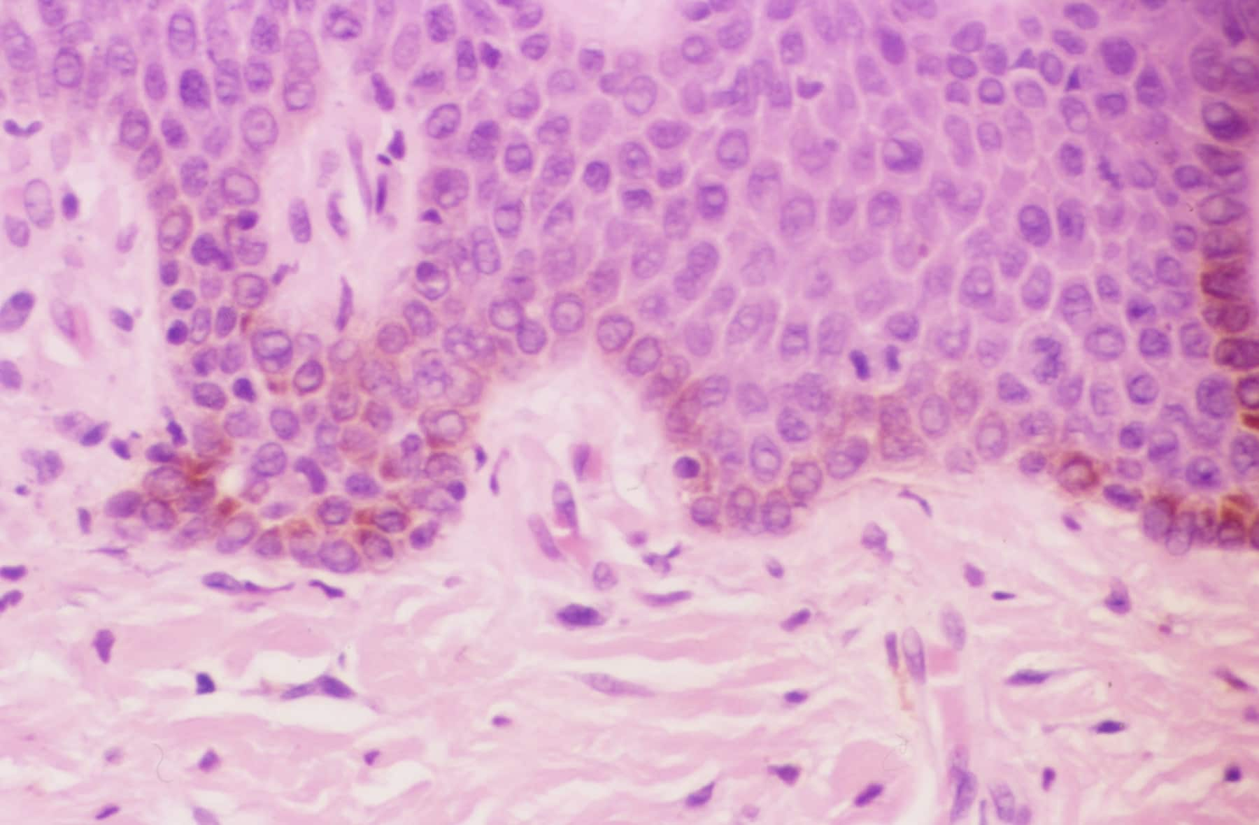
Smoker's melanosis
General
- Benign.
- Seen in ~20% of smokers.[13]
- Presence of find (smoking) dose-dependent, i.e. longer heavier smokers are more likely to have it.
Gross
- Typically labial gingvia or buccal mucosa.[13]
Microscopic
Features:
- Basal melanosis.
- +/-Melanin incontinence.
Image:
Intramucosal melanocytic nevus
General
- Most common oral nevus.
- Essentially an intradermal melanocytic nevus.
Microscopic
Features:
- Symmetrical lesion.
- "Matures" with depth
- Less cellular with depth
- Less nuclear atypia with depth.
- Smaller cells with depth.
- Smaller nests with depth.
- Rare mitoses (superficial).
- No deep mitoses.
- No destruction of surrounding structures.
- No nucleoli.
Sign out
PALATE LESION, PUNCH BIOPSY:
- INTRAMUCOSAL MELANOCYTIC NEVUS.
See also
References
- ↑ 1.0 1.1 Cotran, Ramzi S.; Kumar, Vinay; Fausto, Nelson; Nelso Fausto; Robbins, Stanley L.; Abbas, Abul K. (2005). Robbins and Cotran pathologic basis of disease (7th ed.). St. Louis, Mo: Elsevier Saunders. pp. 777. ISBN 0-7216-0187-1.
- ↑ Kanitakis, J.; Zambruno, G.; Marchand, C.; Perret-Liaudet, P.; Hermier, C.; Thivolet, J. (1990). "[Oral hairy leukoplakia in AIDS. Histologic and ultrastructural study of 8 cases].". Ann Dermatol Venereol 117 (5): 345-53. PMID 2169222.
- ↑ Itin, PH.; Lautenschlager, S. (1997). "Viral lesions of the mouth in HIV-infected patients.". Dermatology 194 (1): 1-7. PMID 9031782.
- ↑ URL: http://www.pathologyoutlines.com/oralcavity.html#hairyleukoplakia.
- ↑ URL: http://www.emedicine.com/asp/dictionary.asp?keyword=hyperkeratosis.
- ↑ URL: http://www.emedicine.com/asp/dictionary.asp?keyword=acanthosis.
- ↑ Mills, Stacey E; Carter, Darryl; Greenson, Joel K; Reuter, Victor E; Stoler, Mark H (2009). Sternberg's Diagnostic Surgical Pathology (5th ed.). Lippincott Williams & Wilkins. pp. 775. ISBN 978-0781779425.
- ↑ URL: http://emedicine.medscape.com/article/1080948-overview#aw2aab6b3. Accessed on: 20 August 2012.
- ↑ 9.0 9.1 9.2 9.3 Thompson, Lester D. R. (2006). Head and Neck Pathology: A Volume in Foundations in Diagnostic Pathology Series (1st ed.). Churchill Livingstone. pp. 240. ISBN 978-0443069604.
- ↑ Segura Saint-Gerons, R.; Ceballos Salobreña, A.; Toro Rojas, M.; Gándara Rey, JM. (Aug 2006). "Oral manifestations of Cowden's disease. Presentation of a clinical case.". Med Oral Patol Oral Cir Bucal 11 (5): E421-4. PMID 16878060.
- ↑ Oliveira, MA.; Medina, JB.; Xavier, FC.; Magalhães, M.; Ortega, KL. (2010). "Cowden syndrome.". Dermatol Online J 16 (1): 7. PMID 20137749.
- ↑ 12.0 12.1 Fernandez-Flores, A. (Jul 2010). "Solitary oral fibromas of the tongue show similar morphologic features to fibrous papule of the face: a study of 31 cases.". Am J Dermatopathol 32 (5): 442-7. doi:10.1097/DAD.0b013e3181c47142. PMID 20421776.
- ↑ 13.0 13.1 13.2 Kauzman, A.; Pavone, M.; Blanas, N.; Bradley, G. (Nov 2004). "Pigmented lesions of the oral cavity: review, differential diagnosis, and case presentations.". J Can Dent Assoc 70 (10): 682-3. PMID 15530266.