Difference between pages "Autoimmune metaplastic atrophic gastritis" and "Chronic sialadenitis"

From Libre Pathology
(Difference between pages)
Jump to navigation Jump to search
(+caption, re-arrange imges)
 
m (vauthors --> authors)
 
Line 1: Line 1:
{{ Infobox diagnosis
'''Chronic sialadenitis''' is a chronic inflammatory process involving a [[salivary glands|salivary gland]].
| Name      = {{PAGENAME}}
 
| Image      = Metaplastic atrophic gastritis - body -- intermed mag.jpg
'''Chronic sailolithiasis''' redirects here.
| Width      = Metaplastic atrophic gastritis - body of stomach. [[H&E stains]].
'''Sialadenitis''' redirects here.  
| Caption    = Atrophic gastritis (body) without appreciable parietal cells. [[H&E stain]].
| Micro      = corpus predominant inflammation - usu. moderate or severe, loss of parietal cells, increased G cells in the antrum
| Subtypes  =
| LMDDx      = [[chronic gastritis]], [[intestinal metaplasia of the stomach]], [[gastric neuroendocrine tumour]]
| Stains    =
| IHC        =
| EM        =
| Molecular  =
| IF        =
| Gross      =
| Grossing  =
| Site      = [[stomach]]
| Assdx      =
| Syndromes  =
| Clinicalhx =
| Signs      =
| Symptoms  =
| Prevalence = uncommon
| Bloodwork  = antibodies to parietal cells & intrinsic factor, [[macrocytic anemia]], increased gastrin level
| Rads      =
| Endoscopy  = erythema - corpus only
| Prognosis  =
| Other      =
| ClinDDx    = diffuse chronic gastritis
}}
'''Autoimmune metaplastic atrophic gastritis''', also '''autoimmune gastritis'''<ref name=pmid16382988>{{Cite journal  | last1 = Chlumská | first1 = A. | last2 = Boudová | first2 = L. | last3 = Benes | first3 = Z. | last4 = Zámecník | first4 = M. | title = Autoimmune gastritis. A clinicopathologic study of 25 cases. | journal = Cesk Patol | volume = 41 | issue = 4 | pages = 137-42 | month = Oct | year = 2005 | doi =  | PMID = 16382988 }}</ref> (abbreviated '''AIG'''), is a rare pathology of the [[stomach]].  It is closely associated with '''pernicious anemia'''.


==General==
==General==
*Pathology: loss of parietal cells, gastric atrophy.
*Occasionally associated with malignancy, e.g. [[adenoid cystic carcinoma]].<ref name=pmid21159490>{{cite journal |author=Hasegawa M, Cheng J, Maruyama S, ''et al.'' |title=Complication of adenoid cystic carcinoma and sialolithiasis in the submandibular gland: report of a case and its etiological background |journal=Int J Oral Maxillofac Surg |volume=40 |issue=6 |pages=647–50 |year=2011 |month=June |pmid=21159490 |doi=10.1016/j.ijom.2010.11.009 |url=}}</ref>
*Lab: classically considered to have [[macrocytic anemia]]; however, normocytic and microcystic more common.<ref name=pmid16239424>{{Cite journal | last1 = Hershko | first1 = C. | last2 = Ronson | first2 = A. | last3 = Souroujon | first3 = M. | last4 = Maschler | first4 = I. | last5 = Heyd | first5 = J. | last6 = Patz | first6 = J. | title = Variable hematologic presentation of autoimmune gastritis: age-related progression from iron deficiency to cobalamin depletion. | journal = Blood | volume = 107 | issue = 4 | pages = 1673-9 | month = Feb | year = 2006 | doi = 10.1182/blood-2005-09-3534 | PMID = 16239424 }}</ref>
*Etiology: autoimmune.


Diagnosis based on serology for antibodies to:<ref name=pmid12643357>{{Cite journal  | last1 = Oh | first1 = R. | last2 = Brown | first2 = DL. | title = Vitamin B12 deficiency. | journal = Am Fam Physician | volume = 67 | issue = 5 | pages = 979-86 | month = Mar | year = 2003 | doi =  | PMID = 12643357 }}</ref>
Etiology:<ref>URL: [http://emedicine.medscape.com/article/882358-overview http://emedicine.medscape.com/article/882358-overviewhttp://emedicine.medscape.com/article/882358-overview]. Accessed on: 10 January 2011.</ref>
*Parietal cells.
*Infection.
*Intrinsic factor.  
*Autoimmune (e.g. [[Sjögren syndrome]], [[systemic lupus erythematosus]]).
*Other.


Others:
Associations:
*Gastrin level (increased).<ref name=pmid21947876>{{Cite journal | last1 = Annibale | first1 = B. | last2 = Lahner | first2 = E. | last3 = Fave | first3 = GD. | title = Diagnosis and management of pernicious anemia. | journal = Curr Gastroenterol Rep | volume = 13 | issue = 6 | pages = 518-24 | month = Dec | year = 2011 | doi = 10.1007/s11894-011-0225-5 | PMID = 21947876 }}</ref>
*[[Smoking]].<ref name=pmid2037973>{{cite journal |author=Eliasson L, Heyden G, Landahl S, Steen B |title=Effects of tobacco and diuretics on human palatal salivary glands |journal=J. Oral Pathol. Med. |volume=20 |issue=3 |pages=126–9 |year=1991 |month=March |pmid=2037973 |doi= |url=}}</ref> (???)
**Normal < 100 pg/mL.<ref>URL: [http://www.mayomedicallaboratories.com/test-catalog/Clinical+and+Interpretive/8512 http://www.mayomedicallaboratories.com/test-catalog/Clinical+and+Interpretive/8512]. Accessed on: 14 August 2012.</ref>
 
Note:
*Parietal cells produce ''intrinsic factor'' (important for vitamin B12 absorption) and ''hydrogen chloride'', i.e. stomach acid.


==Gross==
==Gross==
*Erythema - corpus involved, antrum spared.
Features:
*Typical location: submandibular salivary gland.
*Salivary gland swelling.<ref name=pmid21159490/>


==Microscopic==
==Microscopic==
Features:
Features:
*Corpus predominant inflammation - usu. moderate or severe - '''key feature'''.
*Non-neoplastic mononuclear inflammatory infiltrate (lymphocytes, [[plasma cell]]s).
*Loss of parietal cells.
*Fibrosis.
*Increased G cells in the antrum.
*+/-Calcifications.
**Produce gastrin to stimulate the (missing) parietal cells.


Notes:
Note:
*Compare with other types of ''[[gastric atrophy]]''.
*If the infiltrate is predominantly lymphocytes Sjögren's is a possibility, and reporting a ''[[Focus score]]'' should be considered.


DDx:
DDx:
*[[Gastric neuroendocrine tumour]].
*[[Lymphoma]] - especially [[MALT lymphoma]].<ref name=pmid22475637>{{Cite journal  | last1 = Beasley | first1 = MJ. | title = Lymphoma of the Thyroid and Head and Neck. | journal = Clin Oncol (R Coll Radiol) | volume =  | issue =  | pages =  | month = Apr | year = 2012 | doi = 10.1016/j.clon.2012.02.010 | PMID = 22475637 }}</ref>
*[[Intestinal metaplasia of the stomach]] with chronic inflammation.
*[[IgG4-related systemic diseases|IgG4-related sialadenitis]].<ref name=pmid31760789>{{cite journal |authors=Thompson LDR |title=IgG4-Related Sialadenitis |journal=Ear Nose Throat J |volume= |issue= |pages=145561319890153 |date=November 2019 |pmid=31760789 |doi=10.1177/0145561319890153 |url=}}</ref>


===Images===
===Image===
<gallery>
<gallery>
Image: Metaplastic atrophic gastritis - body -- very low mag.jpg | MAG (body) - very low mag.
Image:Chronic_sialadenitis.jpg | 
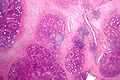
Chronic sialadenitis. (WC/Nephron)
Image: Metaplastic atrophic gastritis - body -- low mag.jpg | MAG (body) - low mag.
Image: Metaplastic atrophic gastritis - body - alt -- low mag.jpg | MAG (body) - low mag.
Image: Metaplastic atrophic gastritis - body -- intermed mag.jpg | MAG (body) - intermed. mag.
Image: Metaplastic atrophic gastritis - body -- high mag.jpg | MAG (body) - high mag.
Image: Metaplastic atrophic gastritis - body - alt -- high mag.jpg | MAG (body) - high mag.
Image: Metaplastic atrophic gastritis - body -- very high mag.jpg | MAG (body) - very high mag.
 
Image: Metaplastic atrophic gastritis - body 2 -- very low mag.jpg | MAG (body) - very low mag.
Image: Metaplastic atrophic gastritis - body 2 -- low mag.jpg | MAG (body) - low mag.
Image: Metaplastic atrophic gastritis - body 2 - alt -- low mag.jpg | MAG (body) - low mag.
Image: Metaplastic atrophic gastritis - body 2 -- intermed mag.jpg | MAG (body) - intermed. mag.
Image: Metaplastic atrophic gastritis - body 2 -- high mag.jpg | MAG (body) - high mag.
Image: Metaplastic atrophic gastritis - body 2 - alt -- high mag.jpg | MAG (body) - high mag.


Image: Metaplastic atrophic gastritis - antrum -- low mag.jpg | MAG - antrum - low mag.
Image: Metaplastic atrophic gastritis - antrum -- intermed mag.jpg | MAG - antrum - low mag.
Image: Metaplastic atrophic gastritis - antrum - alt -- intermed mag.jpg | MAG - antrum - low mag.
 
 
</gallery>
 
==IHC==
Features:<ref name=pmid20975338>{{Cite journal  | last1 = Park | first1 = JY. | last2 = Cornish | first2 = TC. | last3 = Lam-Himlin | first3 = D. | last4 = Shi | first4 = C. | last5 = Montgomery | first5 = E. | title = Gastric lesions in patients with autoimmune metaplastic atrophic gastritis (AMAG) in a tertiary care setting. | journal = Am J Surg Pathol | volume = 34 | issue = 11 | pages = 1591-8 | month = Nov | year = 2010 | doi = 10.1097/PAS.0b013e3181f623af | PMID = 20975338 }}</ref>
*Chromogranin A +ve (demonstrates ''nodular enterochromaffin-like cell hyperplasia'').
*Gastrin -ve (body of stomach).
**+ve in antrum.
 
===Images===
<gallery>
Image: Metaplastic atrophic gastritis - body - chromogranin A -- very low mag.jpg | MAG (body) - chromogranin A - very low mag.
Image: Metaplastic atrophic gastritis - body - chromogranin A -- low mag.jpg | MAG (body) - chromogranin A - low mag.
Image: Metaplastic atrophic gastritis - body - chromogranin A -- intermed mag.jpg | MAG (body) - chromogranin A - intermed. mag.
Image: Metaplastic atrophic gastritis - body - chromogranin A -- high mag.jpg | MAG (body) - chromogranin A - high mag.
 
Image: Metaplastic atrophic gastritis - body - gastrin -- low mag.jpg | MAG (body) - gastrin - low mag.
Image: Metaplastic atrophic gastritis - body - alt - gastrin -- low mag.jpg | MAG (body) - gastrin - low mag.
 
Image: Metaplastic atrophic gastritis - antrum - gastrin -- low mag.jpg | MAG (antrum) - gastrin - low mag.
Image: Metaplastic atrophic gastritis - antrum - gastrin -- intermed mag.jpg | MAG (antrum) - gastrin - intermed. mag.
</gallery>
</gallery>
====www====
*[http://www.ncbi.nlm.nih.gov/pmc/articles/PMC2575912/figure/f5/ Autoimmune gastritis - chromogranin A (nih.gov)].<ref name=pmid18719002>{{Cite journal  | last1 = Pritchard | first1 = DM. | last2 = Berry | first2 = D. | last3 = Przemeck | first3 = SM. | last4 = Campbell | first4 = F. | last5 = Edwards | first5 = SW. | last6 = Varro | first6 = A. | title = Gastrin increases mcl-1 expression in type I gastric carcinoid tumors and a gastric epithelial cell line that expresses the CCK-2 receptor. | journal = Am J Physiol Gastrointest Liver Physiol | volume = 295 | issue = 4 | pages = G798-805 | month = Oct | year = 2008 | doi = 10.1152/ajpgi.00015.2008 | PMID = 18719002 }}</ref>
**Findings may be seen in hypergastrinemia and nodular enterochromaffin cell-like (ECL) hyperplasia.


==Sign out==
==Sign out==
<pre>
<pre>
STOMACH, BIOPSY:
SUBMANDIBULAR GLAND, RIGHT, EXCISION:
- SEVERE CHRONIC ACTIVE GASTRITIS WITH EXTENSIVE INTESTINAL METAPLASIA.
- CHRONIC SIALOADENITIS.
- NEGATIVE FOR HELICOBACTER-LIKE ORGANISMS.
- SIALOLITHIASIS.
- NEGATIVE FOR DYSPLASIA AND NEGATIVE FOR MALIGNANCY.
- TWO BENIGN LYMPH NODES.
- NEGATIVE FOR MALIGNANCY.
</pre>


COMMENT:
====Micro====
Parietal cells are not apparent on the H&E stained sections. Immunostains show  
The sections show submandibular salivary gland with a mild patchy mixed mononuclear cell
rows of Chromogranin A positive cells and a lack of gastrin staining.
infiltrate, fibrosis and a large benign calcification. No zonal necrosis is identified.
Significant nuclear atypia is not identified.


These findings suggest an autoimmune gastritis; correlation with blood work
=====Alternate=====
is suggested.
The sections show a salivary gland with a patchy mixed mononuclear cell infiltrate and fibrosis. Significant nuclear atypia is not identified. Plasma cells are not prominent. Germinal centres are present.
</pre>


==See also==
==See also==
*[[Stomach]].
*[[Salivary gland]].
*[[Gastritis]].
**[[Chronic gastritis]].
*[[Anemia]].


==References==
==References==
{{Reflist|2}}
{{Reflist|2}}


[[Category:Stomach]]
[[Category:Diagnosis]]
[[Category:Diagnosis]]
[[Category:Salivary gland]]

Revision as of 20:28, 24 May 2020

Chronic sialadenitis is a chronic inflammatory process involving a salivary gland.

Chronic sailolithiasis redirects here. Sialadenitis redirects here.

General

Etiology:[2]

Associations:

Gross

Features:

  • Typical location: submandibular salivary gland.
  • Salivary gland swelling.[1]

Microscopic

Features:

  • Non-neoplastic mononuclear inflammatory infiltrate (lymphocytes, plasma cells).
  • Fibrosis.
  • +/-Calcifications.

Note:

  • If the infiltrate is predominantly lymphocytes Sjögren's is a possibility, and reporting a Focus score should be considered.

DDx:

Image

Sign out

SUBMANDIBULAR GLAND, RIGHT, EXCISION:
- CHRONIC SIALOADENITIS.
- SIALOLITHIASIS.
- TWO BENIGN LYMPH NODES.
- NEGATIVE FOR MALIGNANCY.

Micro

The sections show submandibular salivary gland with a mild patchy mixed mononuclear cell infiltrate, fibrosis and a large benign calcification. No zonal necrosis is identified. Significant nuclear atypia is not identified.

Alternate

The sections show a salivary gland with a patchy mixed mononuclear cell infiltrate and fibrosis. Significant nuclear atypia is not identified. Plasma cells are not prominent. Germinal centres are present.

See also

References

  1. 1.0 1.1 Hasegawa M, Cheng J, Maruyama S, et al. (June 2011). "Complication of adenoid cystic carcinoma and sialolithiasis in the submandibular gland: report of a case and its etiological background". Int J Oral Maxillofac Surg 40 (6): 647–50. doi:10.1016/j.ijom.2010.11.009. PMID 21159490.
  2. URL: http://emedicine.medscape.com/article/882358-overviewhttp://emedicine.medscape.com/article/882358-overview. Accessed on: 10 January 2011.
  3. Eliasson L, Heyden G, Landahl S, Steen B (March 1991). "Effects of tobacco and diuretics on human palatal salivary glands". J. Oral Pathol. Med. 20 (3): 126–9. PMID 2037973.
  4. Beasley, MJ. (Apr 2012). "Lymphoma of the Thyroid and Head and Neck.". Clin Oncol (R Coll Radiol). doi:10.1016/j.clon.2012.02.010. PMID 22475637.
  5. Thompson LDR (November 2019). "IgG4-Related Sialadenitis". Ear Nose Throat J: 145561319890153. doi:10.1177/0145561319890153. PMID 31760789.