Difference between revisions of "Odontogenic tumours and cysts"
Jump to navigation
Jump to search
m (→General: +Tx) |
(→Adenomatoid odontogenic tumour: split out) |
||
| (18 intermediate revisions by the same user not shown) | |||
| Line 2: | Line 2: | ||
The general topic of ''head and neck pathology'' is covered in the ''[[head and neck pathology]]'' and ''[[head and neck cytopathology]]'' articles. | The general topic of ''head and neck pathology'' is covered in the ''[[head and neck pathology]]'' and ''[[head and neck cytopathology]]'' articles. | ||
The vast majority of oral malignancies are [[squamous cell carcinoma]]. Common odontogenic cysts are [[dentigerous cyst]]s, and [[radicular cyst]]s.<ref name=pmid20303056>{{Cite journal | last1 = Eichhorn | first1 = W. | last2 = Wehrmann | first2 = M. | last3 = Blessmann | first3 = M. | last4 = Pohlenz | first4 = P. | last5 = Blake | first5 = F. | last6 = Schmelzle | first6 = R. | last7 = Heiland | first7 = M. | title = Metastases in odontogenic cysts: literature review and case presentation. | journal = Oral Surg Oral Med Oral Pathol Oral Radiol Endod | volume = 109 | issue = 4 | pages = 582-6 | month = Apr | year = 2010 | doi = 10.1016/j.tripleo.2009.11.013 | PMID = 20303056 }}</ref> | |||
=Tooth histology 101= | =Tooth histology 101= | ||
| Line 18: | Line 20: | ||
Image: | Image: | ||
<gallery> | |||
Image:Tooth_in_teratoma_-_very_low_mag.jpg | Tooth. (WC) | |||
</gallery> | |||
===Enamel 101=== | ===Enamel 101=== | ||
| Line 68: | Line 72: | ||
===Microscopic=== | ===Microscopic=== | ||
Features: | Features: | ||
*Squamous epithelium - '' | *Squamous epithelium - non-keratinized - '''important'''. | ||
*+/-Giant cells. | *+/-Giant cells. | ||
*+/-Cholesterol clefts. | *+/-Cholesterol clefts. | ||
| Line 91: | Line 95: | ||
===Microscopic=== | ===Microscopic=== | ||
Features: | Features: | ||
*Squamous epithelium - '' | *Squamous epithelium. | ||
**Classically described as non-keratinized - in which case the diagnosis is straight forward - '''important'''. | |||
**Approximately half have keratin.<ref name=pmid9195629>{{Cite journal | last1 = Yoshiura | first1 = K. | last2 = Higuchi | first2 = Y. | last3 = Araki | first3 = K. | last4 = Shinohara | first4 = M. | last5 = Kawazu | first5 = T. | last6 = Yuasa | first6 = K. | last7 = Tabata | first7 = O. | last8 = Kanda | first8 = S. | title = Morphologic analysis of odontogenic cysts with computed tomography. | journal = Oral Surg Oral Med Oral Pathol Oral Radiol Endod | volume = 83 | issue = 6 | pages = 712-8 | month = Jun | year = 1997 | doi = | PMID = 9195629 }}</ref> | |||
*+/-Giant cells. | *+/-Giant cells. | ||
*+/-Cholesterol clefts. | *+/-Cholesterol clefts. | ||
| Line 97: | Line 103: | ||
DDx: | DDx: | ||
*[[Radicular cyst]] - history is the '''key''' to differentiate. | *[[Radicular cyst]] - history is the '''key''' to differentiate. | ||
*[[Keratocystic odontogenic tumour]] - | *[[Keratocystic odontogenic tumour]] - parakeratosis, ribbon like, (artefactual) clefting. | ||
Images: | Images: | ||
*[http://www.surgicalpathologyatlas.com/glfusion/mediagallery/media.php?f=0&sort=0&s=20080802170149187 Dentigerous cyst (surgicalpathologyatlas.com)]. | *[http://www.surgicalpathologyatlas.com/glfusion/mediagallery/media.php?f=0&sort=0&s=20080802170149187 Dentigerous cyst (surgicalpathologyatlas.com)]. | ||
*[http://www.ncbi.nlm.nih.gov/pmc/articles/PMC3180832/figure/F2/ Dentigerous cyst (nih.gov)].<ref name=pmid21957386>{{Cite journal | last1 = Moosvi | first1 = Z. | last2 = Tayaar | first2 = SA. | last3 = Kumar | first3 = GS. | title = Neoplastic potential of odontogenic cysts. | journal = Contemp Clin Dent | volume = 2 | issue = 2 | pages = 106-9 | month = Apr | year = 2011 | doi = 10.4103/0976-237X.83073 | PMID = 21957386 | PMC = 3180832 }}</ref> | *[http://www.ncbi.nlm.nih.gov/pmc/articles/PMC3180832/figure/F2/ Dentigerous cyst (nih.gov)].<ref name=pmid21957386>{{Cite journal | last1 = Moosvi | first1 = Z. | last2 = Tayaar | first2 = SA. | last3 = Kumar | first3 = GS. | title = Neoplastic potential of odontogenic cysts. | journal = Contemp Clin Dent | volume = 2 | issue = 2 | pages = 106-9 | month = Apr | year = 2011 | doi = 10.4103/0976-237X.83073 | PMID = 21957386 | PMC = 3180832 }}</ref> | ||
===Sign out=== | |||
====Keratinized==== | |||
<pre> | |||
MAXILLARY SINUS CYST, LEFT, EXCISION: | |||
- ACANTHOTIC STRATIFIED SQUAMOUS EPITHELIUM WITH INFLAMMATION, COMPACT | |||
KERATIN AND FOCAL PARAKERATOSIS -- CONSISTENT WITH DENTIGEROUS CYST WITH KERATIN. | |||
- BENIGN BONE. | |||
- NEGATIVE FOR ODONTOGENIC KERATOCYSTIC TUMOUR (ODONTOGENIC KERATOCYST). | |||
</pre> | |||
==Keratocystic odontogenic tumour== | ==Keratocystic odontogenic tumour== | ||
{{Main|Keratocystic odontogenic tumour}} | |||
==Ameloblastoma== | ==Ameloblastoma== | ||
{{Main|Ameloblastoma}} | |||
==Adenomatoid odontogenic tumour== | ==Adenomatoid odontogenic tumour== | ||
{{Main|Adenomatoid odontogenic tumour}} | |||
==Ameloblastic fibroma== | ==Ameloblastic fibroma== | ||
| Line 239: | Line 155: | ||
Features: | Features: | ||
*Paucicellular lesion with pale staining. | *Paucicellular lesion with pale staining. | ||
==Squamous odontogenic tumour== | |||
{{Main|Squamous odontogenic tumour}} | |||
=See also= | =See also= | ||
| Line 252: | Line 171: | ||
[[Category:Head and neck pathology]] | [[Category:Head and neck pathology]] | ||
[[Category:Odontogenic tumours and cysts|Odontogenic tumours and cysts]] | |||
Latest revision as of 00:54, 24 March 2019
This article covers odontogenic tumours and cysts, which is a subset of oral pathology and can be grouped under the heading of head and neck pathology.
The general topic of head and neck pathology is covered in the head and neck pathology and head and neck cytopathology articles.
The vast majority of oral malignancies are squamous cell carcinoma. Common odontogenic cysts are dentigerous cysts, and radicular cysts.[1]
Tooth histology 101
Teeth develop from a combination of:
- Epithelium (downward growth).
- Mesenchyme.
Identifying stuff
Pulp:
- Paucicellular.
- Pale staining.
Enamel:
- Hyperchromatic (dark purple).
- "Fish scale" appearance.
Image:
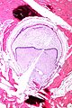
Tooth. (WC)

Enamel 101
- Arises from reduced enamel epithelium.
Reduced enamel epithelium
Microscopic
Features:
- Bilayered epithelium consisting of:
- Cuboidal/columnar cells with:
- Moderate eosinophilic cytoplasm.
- Round (slightly irregular) centrally place nuclei.
- Cuboidal/columnar cells with:
Notes:
- Transforms into squamous epithelium. (???)
Specific entities
Odontoma
General
- Usually diagnosed clinically.
- Benign.
- Most common odontogenic tumour - considered to be a hamartoma.[2]
- Etiology unknown.[3]
- Typically first two decades of life.
Classification:[2]
- Compound odontoma - tooth-like structure.
- Complex odontoma - disorganized mass of odontogenic tissues.
Microscopic
Features:[2]
- Dentin.
- Cementum.
- Pulpal tissue.
- Enamel - has a "fish-scale" appearance.
- Usually lost during decalcificiation.
Images:
Radicular cyst
- AKA periapical cyst.
Clinical
- Non-vital tooth - key feature.
- The tooth that has lost its nerve.
Microscopic
Features:
- Squamous epithelium - non-keratinized - important.
- +/-Giant cells.
- +/-Cholesterol clefts.
- +/-Abundant plasma cells.
DDx:
- Dentigerous cyst - history is the key to differentiate.
- Keratocystic odontogenic tumour - keratinized epithelium.
- Plasma cell neoplasm[4] - should be considered if the lesion is not associated with a carious tooth.
Dentigerous cyst
General
- Unerupted tooth - usually wisdom teeth.
- Young adults.
Treatment:
- Complete removal - as may transform to squamous cell carcinoma or ameloblastoma.[5]
Gross
- Lesion at crown of tooth.
Microscopic
Features:
- Squamous epithelium.
- Classically described as non-keratinized - in which case the diagnosis is straight forward - important.
- Approximately half have keratin.[6]
- +/-Giant cells.
- +/-Cholesterol clefts.
DDx:
- Radicular cyst - history is the key to differentiate.
- Keratocystic odontogenic tumour - parakeratosis, ribbon like, (artefactual) clefting.
Images:
Sign out
Keratinized
MAXILLARY SINUS CYST, LEFT, EXCISION:
- ACANTHOTIC STRATIFIED SQUAMOUS EPITHELIUM WITH INFLAMMATION, COMPACT
KERATIN AND FOCAL PARAKERATOSIS -- CONSISTENT WITH DENTIGEROUS CYST WITH KERATIN.
- BENIGN BONE.
- NEGATIVE FOR ODONTOGENIC KERATOCYSTIC TUMOUR (ODONTOGENIC KERATOCYST).
Keratocystic odontogenic tumour
Main article: Keratocystic odontogenic tumour
Ameloblastoma
Main article: Ameloblastoma
Adenomatoid odontogenic tumour
Main article: Adenomatoid odontogenic tumour
Ameloblastic fibroma
General
- Paedatric population.
Microscopic
Features:
- Palisaded nuclei.
- Fibrous stroma.
Notes:
- No stellate reticulum.
DDx:
Odontogenic myxoma
General
- Benign tumour of mesenchymal origin.
- Often reoccurs.
- Radiologic DDx includes ameloblastoma.
Gross
- Gelatinous mass.
Microscopic
Features:
- Paucicellular lesion with pale staining.
Squamous odontogenic tumour
Main article: Squamous odontogenic tumour
See also
References
- ↑ Eichhorn, W.; Wehrmann, M.; Blessmann, M.; Pohlenz, P.; Blake, F.; Schmelzle, R.; Heiland, M. (Apr 2010). "Metastases in odontogenic cysts: literature review and case presentation.". Oral Surg Oral Med Oral Pathol Oral Radiol Endod 109 (4): 582-6. doi:10.1016/j.tripleo.2009.11.013. PMID 20303056.
- ↑ 2.0 2.1 2.2 2.3 Nelson, BL.; Thompson, LD. (Dec 2010). "Compound odontoma.". Head Neck Pathol 4 (4): 290-1. doi:10.1007/s12105-010-0186-2. PMID 20533004.
- ↑ Yadav, M.; Godge, P.; Meghana, SM.; Kulkarni, SR. (Apr 2012). "Compound odontoma.". Contemp Clin Dent 3 (Suppl 1): S13-5. doi:10.4103/0976-237X.95095. PMID 22629054.
- ↑ Dhanrajani, PJ.; Abdulkarim, SA.. "Multiple myeloma presenting as a periapical lesion in the mandible.". Indian J Dent Res 8 (2): 58-61. PMID 9495138.
- ↑ Kumar, Vinay; Abbas, Abul K.; Fausto, Nelson; Aster, Jon (2009). Robbins and Cotran pathologic basis of disease (8th ed.). Elsevier Saunders. pp. 748. ISBN 978-1416031215.
- ↑ Yoshiura, K.; Higuchi, Y.; Araki, K.; Shinohara, M.; Kawazu, T.; Yuasa, K.; Tabata, O.; Kanda, S. (Jun 1997). "Morphologic analysis of odontogenic cysts with computed tomography.". Oral Surg Oral Med Oral Pathol Oral Radiol Endod 83 (6): 712-8. PMID 9195629.
- ↑ Moosvi, Z.; Tayaar, SA.; Kumar, GS. (Apr 2011). "Neoplastic potential of odontogenic cysts.". Contemp Clin Dent 2 (2): 106-9. doi:10.4103/0976-237X.83073. PMC 3180832. PMID 21957386. https://www.ncbi.nlm.nih.gov/pmc/articles/PMC3180832/.