Difference between revisions of "Nipple adenoma"
(→Images) |
|||
| Line 61: | Line 61: | ||
**Often deeper - one should '''not''' see skin in the histologic section. | **Often deeper - one should '''not''' see skin in the histologic section. | ||
*Syringomatous adenoma | *Syringomatous adenoma | ||
*Intraductal carcinoma | *Intraductal carcinoma - cribriforming glands should be absent | ||
*Invasive ductal carcinoma - IHC is useful (see below) the ducts should all be lined by myoepithelium. | |||
===Images=== | ===Images=== | ||
Revision as of 10:31, 26 March 2015
| Nipple adenoma | |
|---|---|
| Diagnosis in short | |
 Nipple adenoma. H&E stain. | |
|
| |
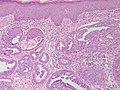
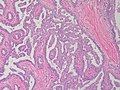
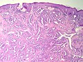
| LM | proliferation of epithelial and myoepithelial elements that extends into the breast stroma; not encapsulated; lacks true fibrovascular cores, +/-focal necrosis |
| LM DDx | intraductal papilloma |
| Site | breast - nipple |
|
| |
| Prevalence | uncommon |
| Prognosis | benign |
| Clin. DDx | Paget's disease of the breast |
Nipple adenoma is a benign pathology of the breast.
It is also known as nipple duct adenoma, nipple adenoma of breast, adenoma of the nipple and florid papillomatosis of the nipple.[1]
General
Clinical DDx:
Microscopic
Features:
- Proliferation of epithelial and myoepithelial elements that extends into the breast stroma.[4]
- Arborising papillomatous epithelial proliferation within duct
- Papillae have fibrovascular cores.
- Florid epithelial hyperplasia can be seen
- Can see haphazard arrangement of proliferating tubular structures
• Differential diagnosis: syringomatous adenoma and tubular carcinoma
Notes:
DDx:
- Intraductal papilloma.
- Found within the duct not the stroma.
- Often deeper - one should not see skin in the histologic section.
- Syringomatous adenoma
- Intraductal carcinoma - cribriforming glands should be absent
- Invasive ductal carcinoma - IHC is useful (see below) the ducts should all be lined by myoepithelium.
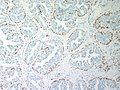
Images
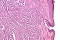
Nipple adenoma - low mag. (WC/Nephron)
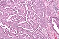
Nipple adenoma - intermed. mag. (WC/Nephron)
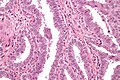
Nipple adenoma - very high mag. (WC/Nephron)
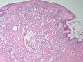
Breast Nipple Adenoma - low power (SKB)
Breast Nipple Adenoma - medium power (SKB)
Breast Nipple Adenoma - medium power (SKB)
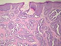
Breast Nipple Adenoma - low power (SKB)
Breast Nipple Adenoma - low power (SKB)
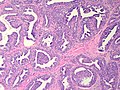
Breast Nipple Adenoma - medium power (SKB)
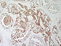
Breast Nipple Adenoma - CK5 (SKB)
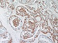
Breast Nipple Adenoma CK14 (SKB)
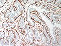
Breast Nipple Adenoma - SMA (SKB)
Breast Nipple Adenoma - p63 (SKB)


www:
See also
References
- ↑ 1.0 1.1 Boutayeb, S.; Benomar, S.; Sbitti, Y.; Harroudi, T.; Hassam, B.; Errihani, H. (2012). "Nipple adenoma in a man: An unusual case report.". Int J Surg Case Rep 3 (5): 190-2. doi:10.1016/j.ijscr.2011.05.008. PMID 22342578.
- ↑ Shinn, L.; Woodward, C.; Boddu, S.; Jha, P.; Fouroutan, H.; Péley, G.. "Nipple adenoma arising in a supernumerary mammary gland: a case report.". Tumori 97 (6): 812-4. doi:10.1700/1018.11102. PMID 22322852.
- ↑ HANDLEY, RS.; THACKRAY, AC. (Jun 1962). "Adenoma of nipple.". Br J Cancer 16: 187-94. PMC 2070922. PMID 13904317. http://www.ncbi.nlm.nih.gov/pmc/articles/PMC2070922/?tool=pubmed.
- ↑ 4.0 4.1 "Adenoma of Nipple.". Br Med J 1 (5330): 563. Mar 1963. PMC 2123505. PMID 20789667. http://www.ncbi.nlm.nih.gov/pmc/articles/PMC2123505/?page=1.
- ↑ URL: http://surgpathcriteria.stanford.edu/breast/nippleadenoma/printable.html. Accessed on: 6 August 2011.
- ↑ Lefkowitch, Jay H. (2006). Anatomic Pathology Board Review (1st ed.). Saunders. pp. 307 Q16. ISBN 978-1416025887.