Difference between revisions of "Neuropathology"
(→Histopathology: more) |
(→Cerebral cortex: more) |
||
| Line 326: | Line 326: | ||
#*Not prominent in frontal cortex. | #*Not prominent in frontal cortex. | ||
#*Where the thalamic axons end. | #*Where the thalamic axons end. | ||
#*Divided in three (''a'', ''b'', ''c'') in the calcarine cortex due to two white matter bands (external band of Baillarger, internal band of Baillarger) than are grossly identified as the ''line of Gennari''.<ref name=Ref_PSNP24>{{Ref PSNP|24}}</ref> | |||
#Inner pyramidal layer. | #Inner pyramidal layer. | ||
#*Location of ''Betz neurons'' - large motor neurons of cerebral cortex. | #*Location of ''Betz neurons'' - large motor neurons of cerebral cortex. | ||
#Polymorphic layer. | #Multiforme layer (Polymorphic layer). | ||
Images: | Images: | ||
Revision as of 05:41, 31 October 2010
Neuropathology is the bane of many anatomical pathologists in teaching hospitals... 'cause they have to fill in for the neuropathologist when he or she is on vacation.
This article is an introduction to neuropathology. There are separate articles for brain tumours, the pituitary gland and muscle pathologies.
Gross
Important
- Uncus (as in uncal herniation).
- Cerebellar tonsils (as in tonsillar herniation).
- Longitudinal fissure - divides cerebrum into hemispheres.
- Lateral sulcus (Sylvian fissure, lateral fissure) - separates temporal lobe from frontal lobe & parietal lobe.
- Central sulcus - separate parietal lobe from frontal lobe.
- Brain stem = medulla oblongata, pons, mesencephalon (midbrain).[1]
Less important
- Glomeruli of Arnold.
- Over lies hippocampus.
- Calcarine cortex - occipital lobe
- Line of Gennari -- very thin white line in the grey matter.
- Image: Calcarine cortex (med.utah.edu) as part of CNS collection (gfmer.ch).
- Nucleus accumbens (NAcc) - inferior-medial to where the internal capsule ends; anterior of optic chiasm.
- Images: NAcc - sagittal (WC), NAcc - frontal (WC).
Trivia
- Claustrum - thin band of grey mater in the external capsule; function uncertain.[2]
Vascular structures
- Posterior cerebellar arteries.
- Inferior of posterior cerebral arteries.
- Anterior inferior cerebellar arteries.
- Branch off basilar artery.
- Posterior inferior cerebellar arteries - AKA PICA.
- Branch off vertebral arteries.
Images:
Meninges
Deep to superficial:
- Pia mater.
- Arachanoid membrane.
- Subarachanoid space - contains blood vessels.
- Dura mater.
- Tough outer covering.
Lesion location classification
Locations:[3]
- Intra-axial = inside the (middle) of spinal cord/brain.
- AKA intramedullary.
- Intradural = not intra-axial, but deep to the dural.
- AKA extramedullary.
- Extradural = outside of dura.
- The above descriptors are often found in radiology reports.
DDx based on location[4]
Intra-axial:
- Glioma (astrocytoma, oligodendroglioma).
- Hemangioblastoma.
- Ependymoma.
Intradural:
- Meningioma.
- Schwannoma.
- Neurofibromas.
- Arachnoid cyst.
Extradural:
- Mets (lung, breast, etc.).
- Schwannoma.
- Sarcoma.
- Plasmacytoma.
- Primary bone tumors - osteosarcoma, osteochondroma, chondrosarcoma.
- Chordoma.
Sampling - sections (autopsy)
- See autopsy.
Standard histologic sections:
| Routine[5] | Head injury[6] | Epilepsy[7] | Dementia[8] | |
| Frontal cortex | Y [1] | Y - bilateral parasagittal [2] | N | Y - middle frontal gyrus [1] |
| Cingulate gyrus | N | N | Y - parasagittal [1] | Y [1] |
| Basal ganglia & internal capsule |
Y [1] | Y - bilateral with corpus callosum [2] | Y - caudate, putamen, globus palidus [1] |
Y - putamen, globus palidus [1] |
| Basal ganglia, internal capsule, thalamus |
N | Y [2] | N | N |
| Temporal lobe | N | N | Y - superior & middle temporal gyri [2] | Y - superior & middle temporal gyri [1] |
| Hippocampus | Y [1] | Y - bilateral [2] | Y - also parahippocampal gyri [2] | Y - also parahippocampal gyri [1] |
| Splenium of corpus callosum | N | Y [1] | N | N |
| Parietal lobe | N | Y - centrum semiovale (unilateral) [1] | N | Y - inferior [1] |
| Occipital cortex | Y [1] | N | N | Y [1] |
| Midbrain | Y [1] | Y [1] | N | Y [1] |
| Cerebellum (with dentate gyrus) |
Y [1] | Y - bilateral [2] | Y - also vermis [2] | Y [1] |
| Pons | N | Y [1] | N | Y [1] |
| Medulla | Y [1] | Y [1] | N | Y [1] |
| Total sections | [7] | [15] | [8] | [11] |
Normal histology
Normal cells
- Neuron:
- Abundant cytoplasm - key feature.
- Often very large cells, with angled edges.
- Prominent nucleolus.
- Nissl substance (granular perinuclear material - rough ER).
- Glial cells.
- Oligodendrocyte.
- Small round nuclei (lymphocyte-like nucleus) - key feature.
- May resemble a fried egg on H&E (clear cytoplasm, central nucleus).
- Astrocyte.
- Irregular non-ovoid nucleus - key feature.
- Nuclei less dense than in oligodendrocyte.
- Close to blood vessels.
- Form blood-brain barrier.
- Cytoplasm normally not visible.
- Image: astrocyte (med.unsw.edu.au) (in endocrine development).
- Microglia - macrophage of the brain (derived from monocyte).
- May be large.
- May have vesicles.
- Rarely seen in normal tissue.
- Oligodendrocyte.
- Ependyma.
- Simple ciliated cuboidal epithelium.
- Image: Ependyma (stonybrookmedicalcenter.org).
Normal cellular constituents in a table
| Key feature | Other features | Image | |
| Neuron | cytoplasm | Nissl substance (prominent RER), "sharp" corners in cell membrane, nucleolus - usu. prominent[9] |
red neurons (WC) |
| Astrocyte | non-ovoid nucleus | no cytoplasm | (unsw.edu) |
| Oligodendrocyte | round small nucleus | peri-nuclear clearing | |
| Microglia | rod-like shape, may have "bent" nucleus |
rarely seen in normal tissue | (ucsf.edu),(vcu.edu) |
Neurons
There are many types of 'em. Broadly, they can be classified as:
- Pyramidal - have a pyramidal shape.
- Dentrites go to molecular layer.
- Axons go to outside of cortex.
- Non-pyramidal.
Motor neurons:
- Coarse Nissl substance - key feature.
- Nissl described as having a tigroid appearance.[10]
- Polygonal shape.
- Send dendrites in all directions.
Image: Motor neuron (stonybrookmedicalcenter.org).
Histology - where
Subependyma
Features:[11]
- Ependyma (simple ciliated cuboidal epithelium).
- Subependymal plate - connective tissue with blood vessels.
Pons
Features:
- Looks like bacon.[12]
- Image: Pons (stonybrookmedicalcenter.org).
Caudate
Features:
- Neurons with adjacent ependymal lining.[13]
- The caudate forms lateral wall of lateral ventricle.
Putamen
Features:
- Histologically identical to the caudate - but not adjacent to a ventricle, i.e. an ependymal lining.
- Striatopallidal fibers AKA pencils of Wilson - bundles of blue fibres (on H&E LFB).
Globus pallidus
Features:
- Histologically distinct from caudate and putamen.
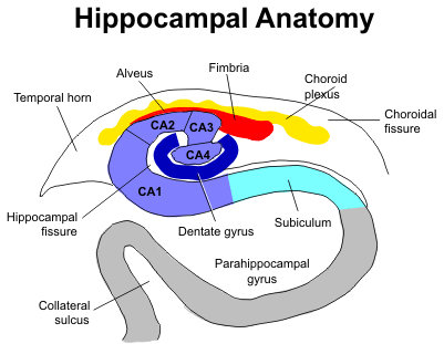
Hippocampus
Hippocampal formation:[14]
- Dentate gyrus.
- "Dense" thin layer of nuclei.
- Quasi "U-shaped"; "open" (top) portion of "U" is superolateral.
- Image: Dentate gyrus (stonybrookmedicalcenter.org).
- Hippocampus proper (AKA Ammon's horn) - this is subdivided:
- CA3 - superior.
- CA1 - inferior (next to subiculum).
- CA2 - in between CA3 and CA1, lateral.
- CA4 - medial (closest to dentate gyrus; CA4 sits in "open" part of "U").
- Subicular complex.
Images:
- Hippocampus - frontal section (WP).
- Hippocampus - good schematic (WC).
- Hippocampus (ajnr.org).
- Hippocampus and subiculum (hu-berlin.de).
- Hippocampus - crappy schematic (ucsd.edu).
Important note:
- CA1 - weak link, dies in ischemia, affected by hypoglycemia.
- CA2 - resistant to ischemia.
DDx of ischemia-like changes in the hippocampus:
- Toxins.
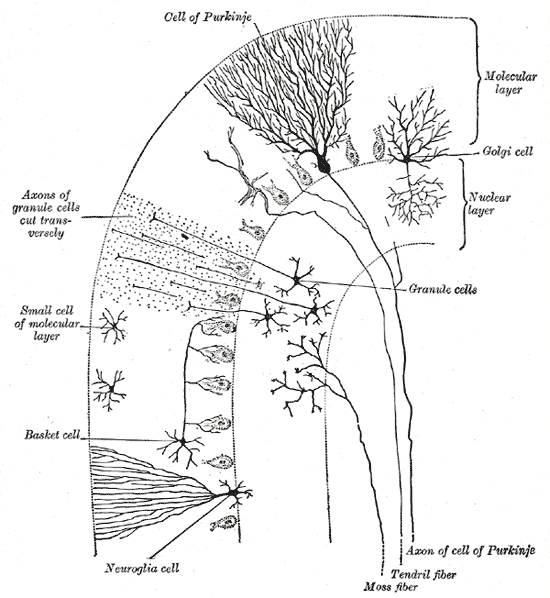
Cerebellum
Main components:
- Cortex (superficial) - branches (Christmas tree-like).
- Dentate nucleus (deep) - looks like the bite impression of a molar.
Cerebellar cortex:
- Layers (superficial to deep) - mnemonic MPG:[15]
- Molecular layer -- "very pink" on H&E.
- Inhibitory interneurons: stellate cells, basket cells.
- Purkinje cell layer.
- One cell layer thick - hueuege cells (~50-80 micrometers[9]).
- Very large nucleus (~4x RBC diameter =~ 4x the size of granule cell).
- Large nucleolus (~1x RBC diameter =~ size of granule cell).
- Very large nucleus (~4x RBC diameter =~ 4x the size of granule cell).
- One cell layer thick - hueuege cells (~50-80 micrometers[9]).
- Granule cell layer -- "very blue" on H&E.
- Granule cells (many), interneurons (Golgi cells --few in number). (???)
- Molecular layer -- "very pink" on H&E.
- Images:
Notes:
- Bergmann glia are found between the molecular layer & granular layer. They are normally not seen. They are increased & prominent in pathologic states (e.g. ischemia); "Bergmann gliosis".[16]
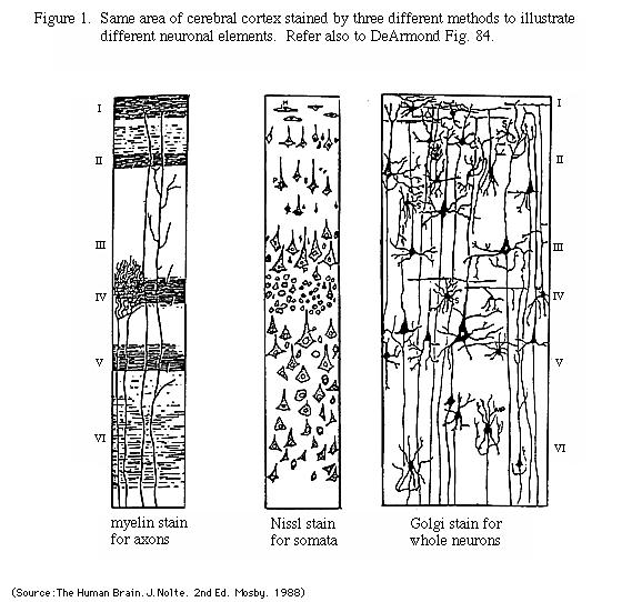
Cerebral cortex
Layers (superficial to deep):
- Molecular layer.
- Empty appearing.
- Outer granular layer.
- Higher cell density & smaller cells than pyramidal layer.
- Outer pyramidal layer.
- Inner granular layer.
- Not prominent in frontal cortex.
- Where the thalamic axons end.
- Divided in three (a, b, c) in the calcarine cortex due to two white matter bands (external band of Baillarger, internal band of Baillarger) than are grossly identified as the line of Gennari.[17]
- Inner pyramidal layer.
- Location of Betz neurons - large motor neurons of cerebral cortex.
- Multiforme layer (Polymorphic layer).
Images:
- Cajal drawings - different areas (WC).
- Different stains (rice.edu).
- Cerebral cortex (williamcalvin.com).
- Cerebral cortex (benbest.com).
Histopathology
Astrocyte changes
Reactive astrocytes.
- Eosinophilic cytoplasm.
- Peripheral nucleus.
- Well-defined cell border.
- Many branching processes.
Types
Alzheimer type II astrocyte:[18]
- Large cleared nucleus - key feature.
- Indistinct cytoplasm.
- Images:
Bergmann gliosis (in the cerebellum):[16]
- Thin layer of cells (2-3 cells), nuclei open and larger than granular cell layer nuclei.
- Image: Bergmann gliosis (sagepub.com).
Creutzfeldt cell:[16]
- Astrocyte that mimics a mitoses; has moderate (identifiable) cytoplasm.
- Finding associated with demyelinating disease.
Other
Axonal swellings:
- Image: Axonal swelling - neuropathologyweb.org.
Architecture
- Rosette = circular/flower-like arrangement of cells[19]
- Pseudorosette = circular/flower-like arrangement of cells with blood vessel at the centre[19]
- Rosenthal fibres = worm-like or corkscrew-like eosinophilic bodies.
- Image: Rosenthal fibres - wikipedia.org.
- Pseudopallisading
Notes: Good set of articles - [20]
Inclusion bodies
- Negri bodies.
- Cytoplasmic inclusions; classically in Purkinje cells of the cerebellum, pyramidal cells of Ammon's horn.
- Rabies.
- Image: Negri bodies (WC/CDC).
- Lewy bodies.
- Eosinophilic cytoplasmic inclusion - composed mostly of alpha-synuclein.[21]
- Image: Lewy body (WC).
Ring enhancing lesions
In HIV/AIDS patients... mass on CT if infection:
- Toxoplasmosis - most common.[22]
Ring enhancing lesion (DDx) - mnemonic MAGICAL DR:[23]
- Metstasis.
- Abscess.
- Glioblastoma.
- Infarct.
- Contusion.
- AIDS-related.
- Lymphoma + HIV assoc. disease (toxoplasma).
- Demyelination (e.g. multiple sclerosis).
- Resolving hematoma.
Alcohol & CNS
Clinical
- Wernicke's encephalopathy
- Mnemonic WACO:
- Wernicke's.
- Ataxia.
- Confusion, confabulation -- Korsakoff.
- Ocular Sx (CN IV palsy).
- Cause: thiamine deficiency.
- Mnemonic WACO:
Pathology
Features:[24]
- Morel's laminar sclerosis = spongy degeneration and gliosis of the cerebral cortex[25] usu. prominent in the third layer of the cortex (outer pyramidal layer) and especially in the lateral-frontal cortex.[26]
- Central pontine myelinolysis.
- Mammillary body shrinkage.[27]
- Anterior cerebellar vermis atrophy; weak finding - as also age-related.[28]
- Vermis atrophy is also seen in schizophrenia.[29]
- Marchiafava-Bignami Disease:
- Rare.
- Demyelination of the corpus callosum.[26]
Common non-specific findings
- Intracranial haemorrhage - due to trauma.
Non-tumour
Acute disseminated encephalomyelitis
General
- Thought to be autoimmune; often associated with/preceded by by viral illness.[30]
- May mimic multiple sclerosis.
- Abbreviated "ADEM".
Diagnosis
- Need to r/o infection (with lumbar puncture).
- No old plaques on imaging (MRI).
Microscopic
Features:
- Spares subcortical fibres (???)
Tx
- Steroids.
- Plasmapheresis.
DDx
- Multiple sclerosis.
Cysts
General
- All are "benign", but some may be fatal due to spatial constraints.
List of cysts
- Colloid cyst.[31]
- Columnar epithelium.
- Arachnoid cyst - considered precursor of meningioma.
- Psammoma bodies.
- Clumps of cells.
- Whorled pattern.
- Dermoid cyst.
- Skin + adnexal structures.
- ... think of ovarian dermoid.
- Epidermoid.
- Choriod cyst.
- ?
- Neuroenteric cyst.
- Epithelial cyst.
Dementia
- Alzheimer's dementia.
- Vascular.
- multi-infarct dementia.
- Parkinson's associated dementia.
- Lewy body dementia.
- Alcohol-related dementia.
- Fronto-temporal dementia (Pick disease).
- Multisystem atrophy.
Mnemonic VITAMIN D VEST:[32]
- Vitamin deficiency (B12, folate, thiamine).
- Infection (HIV).
- Trauma.
- Anoxia.
- Metabolic (Diabetes).
- Intracranial tumour.
- Normal pressure hydrocephalus.
- Degenerative (Alzheimer's, Huntington's, CJD).
- Vascular.
- Endocrine.
- Space occupying lesion (chronic subdural hematoma).
- Toxins (alcohol).
Lewy body dementia
- Parkinsonian features.
- Hallucinations (visual).
- Progressive cog. decline with fluctuations.
Multiple system atrophy
- Alpha-synuclein-rich glial cytoplasmic inclusions - finding at autopsy.[33]
- Alpha-synuclein is implicated in a number of neurodegenerative diseases.[34]
Huntington disease
General
- Autosomal dominant inheritance.
- Mutation: unstable CAG repeat.[35]
Gross
- Missing caudate.[36]
Image: Huntington's disease (ouhsc.edu).
Brain tumours
Tumours are a big part of neuropathology. The most common brain tumour is a metastasis. The most common primary tumour is glioblastoma which has a horrible prognosis.
Paediatric pathology
Joubert syndrome
- Malformation of the cerebellar vermis.[37]
Epidemiology
- Autosomal recessive - mutation in a number of genes including NPHP1, AHI1, and CEP290.[37]
Stroke
Gross
- Soft/mushy brain.
- Older infarcts.
- A "roof" is present - a thin submeningeal layer is preserved by the CSF.[38]
- "Roof" is absent in trauma.
- Cavity - in older infarcts.
- Multiple sclerosis does not cavitate.
- A "roof" is present - a thin submeningeal layer is preserved by the CSF.[38]
- Laminar necrosis = (thin) chalky line replace grey mater.[39]
Hypoxic-ischemic encephalopathy
General
- Often due to cardiac arrest, i.e. global ischemia.
- Triple watershed area = parieto-occipital cortex, extrastriate occipital cortex.
Microscopic
Features:
- Hippocampal ischemic changes:
- Loss of neurons in CA1, CA3 and CA4 +/- "cavitation".
- Neuronal loss: No blue (nuclei) where there should be some.
- Cavitation: bubbles/clear spaces where there should be none.
- CA2 neurons preserved.
- Loss of neurons in CA1, CA3 and CA4 +/- "cavitation".
- "Anoxic neurons".[40]
- Shrunken neurons with intensely eosinophilic cytoplasm and pyknotic (shrunken) nuclei.
- Images:
Multiple sclerosis
General
- A bread 'n butter disease of neurology in Canada.
Radiologic/Gross
Features:[41]
- White matter lesions.
- Cerebrum (classically): periventricular distribution.
- Optic nerves (optic neuritis) - classic presentation.
Microscopic
Features:[42]
- Perivascular inflammation.
- Demyelination.
- Subcortical myelinated fibers are often spared.
Classification of MS lesions:
- Early active.
- Inactive.
- Early remyelinating.
- Late remyelinating.
Weird stuff
Cerebral autosomal dominant arteriopathy with subcortical infarcts and leukoencephalopathy (CADASIL)
General
- Autosomal dominant disorder - the name implies.[43]
- Cases strokes in 40-50 year-old.
- Characteristic MRI findings - present in asymptomatic individuals with mutation.
Etiology
- Mutation of Notch 3 gene.[44]
Diagnosis
- Proven Notch 3 mutation.
- Can be diagnosed on a skin biopsy.
- IHC for Notch 3 -- +ve staining in Notch 3 mutants.
Histology
Features:
- Subcortical infarcts.
- Patches of (non-myelinated) tissue within the white matter deep to the cortex with abundant macrophages.
Note:
- No cortical involvement -- this is unlike multiple sclerosis.
DDx:
- Amyloidosis.
- Binswanger's disease - multi-infarct dementia affecting subcortical white matter.
- Often diagnosed as Alzheimer's disease in the past.
Electron microscopy
- Granular osmiophilic material (GOM).
Binswanger's disease
General
- Multi-infarct dementia affecting subcortical white matter.
- Waste-basket diagnosis; diagnosed if CADASIL and amyloidosis have been excluded.
- Diagnosis has been controversial -- most with this entity (in the past) were diagnosed with Alzheimer's disease.
Microscopic
Features:
- Subcortical lesions that replace the myelin consisting of macrophages.
Prion diseases
Etiology:[45]
- Misfolded cell-surface protein called PrP(C).
Includes:[45]
- Creutzfeldt-Jakob disease (CJD).
- Sporadic fatal insomnia (sFI).
Creutzfeldt-Jakob disease
- Commonly abbreviated as CJD.
- Rare.
- Incurable disease.
- Variant Creutzfeldt-Jakob disease (vCJD).
- Associated with bovine spongiform encephalopathy.
- Should sample: spleen, lymph nodes, tonsils.[46]
Microscopic
Features:
- Spongy appearance (cytoplasmic vacuolization[47]).
Images:
See also
Immunohistochemistry
General
- S-100.
- Sensitive... but non-specific, e.g. also stains melanoma.
Glial
- GFAP (glial fibrillary acidic protein).
Neuronal
- Synaptophysin.
- Chromogranin.
Carcinoma vs. glial tumours
- AE1/AE3 often +ve in glial tumours (e.g. astrocytomas, oligodendrogliomas); CAM5.2 is usu. -ve in glial tumours.[48]
References
- ↑ Martini, Frederic H. (2003). Fundamentals of Anatomy & Physiology (6th ed.). Benjamin Cummings. pp. 466. ISBN 978-0805359336.
- ↑ URL: http://commons.wikimedia.org/wiki/File:Telencephalon-Horiconatal.jpg. Accessed on: 22 September 2010.
- ↑ URL: http://www.nervous-system-diseases.com/spine-tumor.html. Accessed on: 21 September 2010.
- ↑ URL: http://www.nervous-system-diseases.com/spine-tumor.html. Accessed on: 21 September 2010.
- ↑ Burton, Julian L.; Rutty, Guy N. (2010). The Hospital Autopsy A Manual of Fundamental Autopsy Practice (3rd ed.). Oxford University Press. pp. 164. ISBN 978-0340965146. }}
- ↑ Burton, Julian L.; Rutty, Guy N. (2010). The Hospital Autopsy A Manual of Fundamental Autopsy Practice (3rd ed.). Oxford University Press. pp. 176. ISBN 978-0340965146. }}
- ↑ Burton, Julian L.; Rutty, Guy N. (2010). The Hospital Autopsy A Manual of Fundamental Autopsy Practice (3rd ed.). Oxford University Press. pp. 179. ISBN 978-0340965146. }}
- ↑ Burton, Julian L.; Rutty, Guy N. (2010). The Hospital Autopsy A Manual of Fundamental Autopsy Practice (3rd ed.). Oxford University Press. pp. 180. ISBN 978-0340965146. }}
- ↑ 9.0 9.1 Perry, Arie; Brat, Daniel J. (2010). Practical Surgical Neuropathology: A Diagnostic Approach: A Volume in the Pattern Recognition series (1st ed.). Churchill Livingstone. pp. 16. ISBN 978-0443069826.
Cite error: Invalid
<ref>tag; name "Ref_PSNP16" defined multiple times with different content - ↑ URL: http://www.stonybrookmedicalcenter.org/pathology/neuropathology/chapter1. Accessed on: 5 July 2010.
- ↑ Half-day. 28 June 2010.
- ↑ Half-day. 28 June 2010.
- ↑ URL: http://www.stonybrookmedicalcenter.org/pathology/neuropathology/chapter1. Accessed on: 2 July 2010.
- ↑ URL: http://www.stonybrookmedicalcenter.org/pathology/neuropathology/chapter1. Accessed on: 2 July 2010.
- ↑ URL: http://www.stonybrookmedicalcenter.org/pathology/neuropathology/chapter1. Accessed on: 2 July 2010.
- ↑ 16.0 16.1 16.2 Perry, Arie; Brat, Daniel J. (2010). Practical Surgical Neuropathology: A Diagnostic Approach: A Volume in the Pattern Recognition series (1st ed.). Churchill Livingstone. pp. 18. ISBN 978-0443069826.
- ↑ Perry, Arie; Brat, Daniel J. (2010). Practical Surgical Neuropathology: A Diagnostic Approach: A Volume in the Pattern Recognition series (1st ed.). Churchill Livingstone. pp. 24. ISBN 978-0443069826.
- ↑ URL: http://www.neuropathologyweb.org/chapter1/chapter1bAstrocytes.html. Accessed on: 2 July 2010.
- ↑ 19.0 19.1 PMID 16551982
- ↑ http://www.ncbi.nlm.nih.gov/entrez/query.fcgi?cmd=PureSearch&db=PubMed&details_term=Neuropathology%20for%20the%20neuroradiologist
- ↑ Marui W, Iseki E, Kato M, Akatsu H, Kosaka K (August 2004). "Pathological entity of dementia with Lewy bodies and its differentiation from Alzheimer's disease". Acta Neuropathol. 108 (2): 121–8. doi:10.1007/s00401-004-0869-4. PMID 15235805.
- ↑ MUN. Feb 3, 2009.
- ↑ TN2005 NS7.
- ↑ http://www.journals.elsevierhealth.com/periodicals/ycdip/article/S0968-6053(07)00035-X/abstract
- ↑ URL: http://content.karger.com/ProdukteDB/produkte.asp?Doi=114939. Accessed on: 22 September 2010.
- ↑ 26.0 26.1 Johkura K, Naito M, Naka T (March 2005). "Cortical involvement in Marchiafava-Bignami disease". AJNR Am J Neuroradiol 26 (3): 670–3. PMID 15760886. http://www.ajnr.org/cgi/content/full/26/3/670.
- ↑ Shear PK, Sullivan EV, Lane B, Pfefferbaum A (November 1996). "Mammillary body and cerebellar shrinkage in chronic alcoholics with and without amnesia". Alcohol. Clin. Exp. Res. 20 (8): 1489-95. PMID 8947329. http://www3.interscience.wiley.com/resolve/openurl?genre=article&sid=nlm:pubmed&issn=0145-6008&date=1996&volume=20&issue=8&spage=1489.
- ↑ Torvik A (1987). "Brain lesions in alcoholics: neuropathological observations". Acta Med. Scand. Suppl. 717: 47–54. PMID 3478969.
- ↑ Sandyk R, Kay SR, Merriam AE (April 1991). "Atrophy of the cerebellar vermis: relevance to the symptoms of schizophrenia". Int. J. Neurosci. 57 (3-4): 205–12. PMID 1938163.
- ↑ Tenembaum S, Chitnis T, Ness J, Hahn JS (April 2007). "Acute disseminated encephalomyelitis". Neurology 68 (16 Suppl 2): S23–36. doi:10.1212/01.wnl.0000259404.51352.7f. PMID 17438235.
- ↑ MUN. 11 Mar 2009.
- ↑ TN06 PS19
- ↑ Wenning, GK.; Stefanova, N.; Jellinger, KA.; Poewe, W.; Schlossmacher, MG. (Sep 2008). "Multiple system atrophy: a primary oligodendrogliopathy.". Ann Neurol 64 (3): 239-46. doi:10.1002/ana.21465. PMID 18825660.
- ↑ Uversky, VN. (Oct 2008). "Alpha-synuclein misfolding and neurodegenerative diseases.". Curr Protein Pept Sci 9 (5): 507-40. PMID 18855701.
- ↑ Kumar P, Kalonia H, Kumar A (2010). "Huntington's disease: pathogenesis to animal models". Pharmacol Rep 62 (1): 1–14. PMID 20360611.
- ↑ URL: http://moon.ouhsc.edu/kfung/jty1/NeuroTest/Q07-Ans.htm. Accessed on: 29 October 2010.
- ↑ 37.0 37.1 http://www.ninds.nih.gov/disorders/joubert/joubert.htm
- ↑ MUN. 16 December 2009.
- ↑ URL: http://moon.ouhsc.edu/kfung/jty1/neurotest/Q03-Ans.htm. Accessed on: 26 October 2010.
- ↑ URL: http://www.neuropathologyweb.org/chapter2/chapter2aHIE.html. Accessed on: 12 July 2010.
- ↑ URL: http://library.med.utah.edu/kw/ms/path.html. Accessed on: 12 July 2010.
- ↑ URL: http://library.med.utah.edu/kw/ms/path.html. Accessed on: 12 July 2010.
- ↑ Tikka, S.; Mykkänen, K.; Ruchoux, MM.; Bergholm, R.; Junna, M.; Pöyhönen, M.; Yki-Järvinen, H.; Joutel, A. et al. (Apr 2009). "Congruence between NOTCH3 mutations and GOM in 131 CADASIL patients.". Brain 132 (Pt 4): 933-9. doi:10.1093/brain/awn364. PMID 19174371.
- ↑ Kalaria, RN.; Viitanen, M.; Kalimo, H.; Dichgans, M.; Tabira, T. (Nov 2004). "The pathogenesis of CADASIL: an update.". J Neurol Sci 226 (1-2): 35-9. doi:10.1016/j.jns.2004.09.008. PMID 15537516.
- ↑ 45.0 45.1 Watts JC, Balachandran A, Westaway D (March 2006). "The expanding universe of prion diseases". PLoS Pathog. 2 (3): e26. doi:10.1371/journal.ppat.0020026. PMC 1434791. PMID 16609731. https://www.ncbi.nlm.nih.gov/pmc/articles/PMC1434791/.
- ↑ Burton, Julian L.; Rutty, Guy N. (2010). The Hospital Autopsy A Manual of Fundamental Autopsy Practice (3rd ed.). Oxford University Press. pp. 83. ISBN 978-0340965146.
- ↑ URL: http://moon.ouhsc.edu/kfung/jty1/opaq/PathQuiz/N0I002-PQ01-M.htm. Accessed on: 19 October 2010.
- ↑ Perry, Arie; Brat, Daniel J. (2010). Practical Surgical Neuropathology: A Diagnostic Approach: A Volume in the Pattern Recognition series (1st ed.). Churchill Livingstone. pp. 12. ISBN 978-0443069826.