Neurocytoma
Jump to navigation
Jump to search
The printable version is no longer supported and may have rendering errors. Please update your browser bookmarks and please use the default browser print function instead.
Neurocytoma is a rare neuropathology tumour.
General
- Rare.
Microscopic
Features:[1]
- Pineocytomatous/neurocytic rosette = irregular rosette with a large meshwork of fibers (neuropil) at the centre.[2]
- Similar to Homer-Wright rosette.
- Monomorphic cells with round nuclei and speckled chromatin.
- Perinuclear clearing.
- Ganglion cell differentiation.
- Well-defined cell borders.
- Hyalinized vessels.
- Necrosis absent.
IHC
- MIB-1: Usu. low.
- Synapthophysin +ve.
DDx:
- Oligodendroglioma - do not have the characteristic rosettes.
- Ganglioglioma.
- Ependymoma.
- Pilocytic astrocytoma (with predominantly oligo-like cell component).
- Diffuse leptomeningeal gliomeuronal tumour.
Images
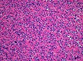
Neurocytoma (H&E: WC, Marvin101).
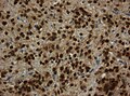
Nuclear NeuN immunoreactivity (WC, Marvin101):


www:
- Neurocytoma (ouhsc.edu).
- Neurocytoma - several images (upmc.edu).
- Neurocytoma - cerebellar - several images (upmc.edu).
IHC
- Synaptophysin +ve.
- Most glial tumour -ve.[3]
Molecular
- FGFR1-TACC1 fusions common.[4]
See also
References
- ↑ URL: http://moon.ouhsc.edu/kfung/jty1/Composites/FNA0IE14-Neurocytoma-Micro.htm. Accessed on: 12 October 2011.
- ↑ Wippold FJ, Perry A (March 2006). "Neuropathology for the neuroradiologist: rosettes and pseudorosettes". AJNR Am J Neuroradiol 27 (3): 488–92. PMID 16551982.
- ↑ URL: http://path.upmc.edu/cases/case383/dx.html. Accessed on: 15 January 2012.
- ↑ Sievers, P.; Stichel, D.; Schrimpf, D.; Sahm, F.; Koelsche, C.; Reuss, DE.; Wefers, AK.; Reinhardt, A. et al. (Jul 2018). "FGFR1:TACC1 fusion is a frequent event in molecularly defined extraventricular neurocytoma.". Acta Neuropathol. doi:10.1007/s00401-018-1882-3. PMID 29978331.