Difference between revisions of "Myoepithelioma"
Jump to navigation
Jump to search
(→IHC) |
m (→General) |
||
| Line 12: | Line 12: | ||
Notes: | Notes: | ||
*First described in 1972.<ref name=pmid5075358>{{Cite journal | last1 = Saksela | first1 = E. | last2 = Tarkkanen | first2 = J. | last3 = Wartiovaara | first3 = J. | title = Parotid clear-cell adenoma of possible myoepithelial origin. | journal = Cancer | volume = 30 | issue = 3 | pages = 742-8 | month = Sep | year = 1972 | doi = | PMID = 5075358 }}</ref> | *First described in 1972.<ref name=pmid5075358>{{Cite journal | last1 = Saksela | first1 = E. | last2 = Tarkkanen | first2 = J. | last3 = Wartiovaara | first3 = J. | title = Parotid clear-cell adenoma of possible myoepithelial origin. | journal = Cancer | volume = 30 | issue = 3 | pages = 742-8 | month = Sep | year = 1972 | doi = | PMID = 5075358 }}</ref> | ||
*May be seen in the skin.<ref name=pmid11224605>{{Cite journal | last1 = Kutzner | first1 = H. | last2 = Mentzel | first2 = T. | last3 = Kaddu | first3 = S. | last4 = Soares | first4 = LM. | last5 = Sangueza | first5 = OP. | last6 = Requena | first6 = L. | title = Cutaneous myoepithelioma: an under-recognized cutaneous neoplasm composed of myoepithelial cells. | journal = Am J Surg Pathol | volume = 25 | issue = 3 | pages = 348-55 | month = Mar | year = 2001 | doi = | PMID = 11224605 }}</ref> | *May be seen in the [[skin]].<ref name=pmid11224605>{{Cite journal | last1 = Kutzner | first1 = H. | last2 = Mentzel | first2 = T. | last3 = Kaddu | first3 = S. | last4 = Soares | first4 = LM. | last5 = Sangueza | first5 = OP. | last6 = Requena | first6 = L. | title = Cutaneous myoepithelioma: an under-recognized cutaneous neoplasm composed of myoepithelial cells. | journal = Am J Surg Pathol | volume = 25 | issue = 3 | pages = 348-55 | month = Mar | year = 2001 | doi = | PMID = 11224605 }}</ref> | ||
==Microsopic== | ==Microsopic== | ||
Features:<ref name=Ref_DCHH130>{{Ref DCHH|130}}</ref> | Features:<ref name=Ref_DCHH130>{{Ref DCHH|130}}</ref> | ||
Revision as of 17:40, 18 March 2015
Myoepithelioma is a benign tumour of the head and neck.
General
- Usually benign.
- May be malignant.
Location - head and neck:[1]
- Parotid gland ~50%.
- Palate ~25%
- Submandibular gland ~12%.
Notes:
Microsopic
Features:[4]
- Myoepithelial cells - may be:
- Spindled.
- Plasmacytoid.
- Epithelioid.
- Clear (rare).
- Lack tubules, i.e. epithelial component.
- May be up to 10% (or 5%[5]).
DDx:
Images
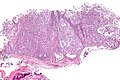
Myoepithelioma - low mag. (WC/Nephron)
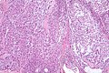
Myoepithelioma - intermed. mag. (WC/Nephron)
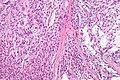
Myoepithelioma - high mag. (WC/Nephron)
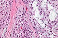
Myoepithelioma - very high mag. (WC/Nephron)


IHC
Features:[4]
- S100 +ve.
- GFAP +ve.
- CK14 +ve.
- Ki-67 low.
Others:[6]
- SMA +ve.
- Calponin +ve.
See also
References
- ↑ Barnes, L.; Appel, BN.; Perez, H.; El-Attar, AM. (Jan 1985). "Myoepithelioma of the head and neck: case report and review.". J Surg Oncol 28 (1): 21-8. PMID 2982059.
- ↑ Saksela, E.; Tarkkanen, J.; Wartiovaara, J. (Sep 1972). "Parotid clear-cell adenoma of possible myoepithelial origin.". Cancer 30 (3): 742-8. PMID 5075358.
- ↑ Kutzner, H.; Mentzel, T.; Kaddu, S.; Soares, LM.; Sangueza, OP.; Requena, L. (Mar 2001). "Cutaneous myoepithelioma: an under-recognized cutaneous neoplasm composed of myoepithelial cells.". Am J Surg Pathol 25 (3): 348-55. PMID 11224605.
- ↑ 4.0 4.1 Tadrous, Paul.J. Diagnostic Criteria Handbook in Histopathology: A Surgical Pathology Vade Mecum (1st ed.). Wiley. pp. 130. ISBN 978-0470519035.
- ↑ I. Weinreb. 24 October 2011.
- ↑ Tadrous, Paul.J. Diagnostic Criteria Handbook in Histopathology: A Surgical Pathology Vade Mecum (1st ed.). Wiley. pp. 18. ISBN 978-0470519035.