Difference between revisions of "Mixed germ cell tumour"
Jump to navigation
Jump to search
(fix) |
|||
| Line 65: | Line 65: | ||
*[[Ovarian tumours]]. | *[[Ovarian tumours]]. | ||
<!-- | |||
==References== | ==References== | ||
{{Reflist|1}} | {{Reflist|1}} | ||
--> | |||
[[Category:Diagnosis]] | [[Category:Diagnosis]] | ||
[[Category:Germ cell tumours]] | [[Category:Germ cell tumours]] | ||
[[Category:Genitourinary pathology]] | [[Category:Genitourinary pathology]] | ||
[[Category:Gynecologic pathology]] | [[Category:Gynecologic pathology]] | ||
Revision as of 09:58, 20 July 2013
| Mixed germ cell tumour | |
|---|---|
| Diagnosis in short | |
 Mixed germ cell tumour. H&E stain. | |
|
| |
| LM | depends on the components |
| LM DDx | other germ cell tumours |
| IHC | variable |
| Site | ovary, testis, mediastinum, other |
|
| |
| Signs | mass lesion |
Mixed germ cell tumour, abbreviated MGCT, is a lesion composed of different germ cell tumours. Most germ cell tumours are mixed.
General
- 60% of GCTs are mixed.
Common combinations:
- Teratoma + embryonal carcinoma + endodermal sinus tumour (yolk sac tumour) (TEE).
- Seminoma + embryonal (SE).
- Teratoma + embryonal +(TE).
Memory device: TEE + all combinations have embryonal carcinoma.
Microscopic
Features:
- Depends on components.
Notes:
- If one cannot identify the component... it is probably yolk sac as this has so many different patterns.
Images
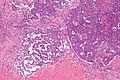
Mixed GCT - intermed mag. (WC/Nephron)
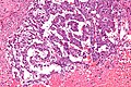
Mixed GCT - high mag. (WC/Nephron)

www:
- Mixed germ cell tumour - several images (upmc.edu).
- Mixed germ cell tumour - several cases (upmc.edu).
IHC
- Beta-hCG +ve - if syncytiotrophoblasts are present.
- AFP +ve - a yolk sac tumour component is present.
- GFAP +ve - if neuroepithelium is present.