Micropapillary urothelial carcinoma
Jump to navigation
Jump to search
| Micropapillary urothelial carcinoma | |
|---|---|
| Diagnosis in short | |
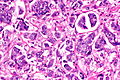
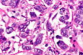
 Urothelial carcinoma with micropapillary features. H&E stain. | |
| Subtypes | (subtype of urothelial carcinoma) |
| LM DDx | conventional urothelial carcinoma, other micropapillary carcinomas (metastases) |
| IHC | CK7 +ve, CK20 +ve/-ve, GATA3 +ve, p63 -ve/+ve |
| Site | urothelium - urinary bladder, ureter, renal pelvis, urethra (males) |
|
| |
| Signs | hematuria (typical presentation) |
| Prevalence | rare |
| Prognosis | poor (aggressive course) |
Micropapillary urothelial carcinoma is an aggressive variant of urothelial carcinoma.[1]
General
Microscopic
Features:[1]
- Micropapillae.
- Quantity of micropapillary (percentage) is variable.[2]
- Conventional urothelial carcinoma (typical).
DDx:
- Metastasis (breast, ovary, lung, pancreas, salivary gland).
Images
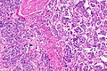
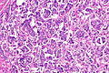
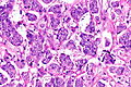
Case
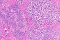
UCCMPF - low mag.
UCCMPF - low mag.
UCCMPF - intermed. mag.
UCCMPF - high mag.
UCCMPF - high mag.
UCCMPF - very high mag.


www
IHC
Features:
Others:[3]
- p63 -ve/+ve.
- p40 -ve/+ve.
Sign out
- Report percentage of micropapillary pattern - suggested.[citation needed]
See also
References
- ↑ 1.0 1.1 1.2 Compérat, E.; Roupret, M.; Yaxley, J.; Reynolds, J.; Varinot, J.; Ouzaïd, I.; Cussenot, O.; Samaratunga, H. (Dec 2010). "Micropapillary urothelial carcinoma of the urinary bladder: a clinicopathological analysis of 72 cases.". Pathology 42 (7): 650-4. doi:10.3109/00313025.2010.522173. PMID 21080874.
- ↑ 2.0 2.1 2.2 2.3 2.4 2.5 2.6 Chatterjee, D.; Das, A.; Radotra, BD.. "Invasive micropapillary carcinoma of urinary bladder: a clinicopathological study.". Indian J Pathol Microbiol 58 (1): 2-6. doi:10.4103/0377-4929.151153. PMID 25673582.
- ↑ 3.0 3.1 Lin, X.; Zhu, B.; Villa, C.; Zhong, M.; Kundu, S.; Rohan, SM.; Yang, XJ. (Sep 2014). "The utility of p63, p40, and GATA-binding protein 3 immunohistochemistry in diagnosing micropapillary urothelial carcinoma.". Hum Pathol 45 (9): 1824-9. doi:10.1016/j.humpath.2014.04.015. PMID 24993315.