Difference between revisions of "Hypertrophic decidual vasculopathy"
Jump to navigation
Jump to search
(+cat.) |
(split out) |
||
| Line 1: | Line 1: | ||
# | '''Hypertrophic decidual vasculopathy''', also known as '''decidual vasculopathy''', is a pathology of the placenta seen in the context of (gestational) [[hypertension]]. | ||
==General== | |||
*A change seen in [[hypertension]]. | |||
*Seen in [[intrauterine growth restriction]] (IUGR). | |||
==Microscopic== | |||
Features:<ref name=pmid18641412>{{Cite journal | last1 = Roberts | first1 = DJ. | last2 = Post | first2 = MD. | title = The placenta in pre-eclampsia and intrauterine growth restriction. | journal = J Clin Pathol | volume = 61 | issue = 12 | pages = 1254-60 | month = Dec | year = 2008 | doi = 10.1136/jcp.2008.055236 | PMID = 18641412 }}</ref> | |||
*Mild or moderate: | |||
*#Perivascular inflammatory cells. | |||
*#+/-Vascular [[thrombosis]]. | |||
*#Smooth muscle hypertrophy. | |||
*#Endothelial hyperplasia. | |||
*#*Above two lead to narrowing of the decidual spiral arteries<ref>AFIP - Placental Pathology. P.122. ISBN: 1-881041-89-1. 2004.</ref> -- '''key feature'''. | |||
*Severe:<ref name=pmid18641412/> | |||
*#Atherosis of maternal blood vessels. | |||
*#*Foamy macrophages within vascular wall. | |||
*#[[Fibrinoid necrosis]] of vessel wall (amorphous eosinophilic material vessel wall). | |||
*Suggestive:<ref>{{Ref Placenta|339}}</ref> | |||
**Decidual vasculitis - lymphocyte predominant without plasma cells. | |||
Note: | |||
*''Smooth muscle hypertrophy'' can also be understood as ''lack of physiological conversion of spiral arteries of the uterus''.<ref name=pmid12848643>{{Cite journal | last1 = Naicker | first1 = T. | last2 = Khedun | first2 = SM. | last3 = Moodley | first3 = J. | last4 = Pijnenborg | first4 = R. | title = Quantitative analysis of trophoblast invasion in preeclampsia. | journal = Acta Obstet Gynecol Scand | volume = 82 | issue = 8 | pages = 722-9 | month = Aug | year = 2003 | doi = | PMID = 12848643 }}</ref> | |||
===Images=== | |||
<gallery> | |||
Image:Hypertrophic_decidual_vasculopathy_intermed_mag.jpg | HDV - intermed. mag. (WC) | |||
Image:Hypertrophic_decidual_vasculopathy_low_mag.jpg | HDV - low mag. (WC) | |||
</gallery> | |||
www: | |||
*[http://path.upmc.edu/cases/case75/images/micro2.jpg Atherosis (upmc.edu)].<ref>URL: [http://path.upmc.edu/cases/case75.html http://path.upmc.edu/cases/case75.html]. Accessed on: 2 January 2012.</ref> | |||
*[http://www.surgpath4u.com/caseviewer.php?case_no=490 Decidual vasculopathy (surgpath4u.com)]. | |||
*[http://www.brown.edu/Courses/Digital_Path/systemic_path/cardio/decidualvasculopathy.html Decidual vasculopathy (brown.edu)]. | |||
==Sign out== | |||
<pre> | |||
PLACENTA, UMBILICAL CORD AND FETAL MEMBRANES, CESAREAN SECTION: | |||
- DECIDUAL VASCULOPATHY. | |||
- PLACENTA SMALL FOR GESTATIONAL AGE (222 GRAMS). | |||
- PLACENTAL DISC WITH EARLY THIRD TRIMESTER VILLI WITH: | |||
-- MULTIPLE PLACENTAL INFARCTS. | |||
-- PERIVILLOUS FIBRIN DEPOSITION. | |||
- THREE VESSEL UMBILICAL CORD WITHIN NORMAL LIMITS. | |||
- FETAL MEMBRANES WITHIN NORMAL LIMITS. | |||
COMMENT: | |||
The 10th percentile placental mass (pre-fixation) for 32 weeks and 6 | |||
days is approximately 247 grams. | |||
</pre> | |||
===Suggestive of decidual vasculopathy=== | |||
<pre> | |||
PLACENTA, UMBILICAL CORD AND FETAL MEMBRANES, CESAREAN SECTION: | |||
- CHANGES SUGGESTIVE OF DECIDUAL VASCULOPATHY (DECIDUAL VASCULITIS). | |||
- PLACENTAL DISC WITH EARLY THIRD TRIMESTER VILLI AND A PLACENTAL INFARCT | |||
(2.5 CM IN MAXIMAL DIMENSION). | |||
- THREE VESSEL UMBILICAL CORD WITHIN NORMAL LIMITS. | |||
- FETAL MEMBRANES WITHIN NORMAL LIMITS. | |||
</pre> | |||
==See also== | |||
*[[Placenta]]. | |||
==References== | |||
{{Reflist|2}} | |||
[[Category:Diagnosis]] | [[Category:Diagnosis]] | ||
[[Category:Placenta]] | |||
Revision as of 07:13, 13 February 2015
Hypertrophic decidual vasculopathy, also known as decidual vasculopathy, is a pathology of the placenta seen in the context of (gestational) hypertension.
General
- A change seen in hypertension.
- Seen in intrauterine growth restriction (IUGR).
Microscopic
Features:[1]
- Mild or moderate:
- Perivascular inflammatory cells.
- +/-Vascular thrombosis.
- Smooth muscle hypertrophy.
- Endothelial hyperplasia.
- Above two lead to narrowing of the decidual spiral arteries[2] -- key feature.
- Severe:[1]
- Atherosis of maternal blood vessels.
- Foamy macrophages within vascular wall.
- Fibrinoid necrosis of vessel wall (amorphous eosinophilic material vessel wall).
- Atherosis of maternal blood vessels.
- Suggestive:[3]
- Decidual vasculitis - lymphocyte predominant without plasma cells.
Note:
- Smooth muscle hypertrophy can also be understood as lack of physiological conversion of spiral arteries of the uterus.[4]
Images
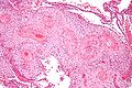
HDV - intermed. mag. (WC)
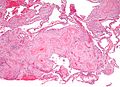
HDV - low mag. (WC)


www:
Sign out
PLACENTA, UMBILICAL CORD AND FETAL MEMBRANES, CESAREAN SECTION:
- DECIDUAL VASCULOPATHY.
- PLACENTA SMALL FOR GESTATIONAL AGE (222 GRAMS).
- PLACENTAL DISC WITH EARLY THIRD TRIMESTER VILLI WITH:
-- MULTIPLE PLACENTAL INFARCTS.
-- PERIVILLOUS FIBRIN DEPOSITION.
- THREE VESSEL UMBILICAL CORD WITHIN NORMAL LIMITS.
- FETAL MEMBRANES WITHIN NORMAL LIMITS.

COMMENT:
The 10th percentile placental mass (pre-fixation) for 32 weeks and 6
days is approximately 247 grams.
Suggestive of decidual vasculopathy
PLACENTA, UMBILICAL CORD AND FETAL MEMBRANES, CESAREAN SECTION:
- CHANGES SUGGESTIVE OF DECIDUAL VASCULOPATHY (DECIDUAL VASCULITIS).
- PLACENTAL DISC WITH EARLY THIRD TRIMESTER VILLI AND A PLACENTAL INFARCT
  (2.5 CM IN MAXIMAL DIMENSION).
- THREE VESSEL UMBILICAL CORD WITHIN NORMAL LIMITS.
- FETAL MEMBRANES WITHIN NORMAL LIMITS.
See also
References
- ↑ 1.0 1.1 Roberts, DJ.; Post, MD. (Dec 2008). "The placenta in pre-eclampsia and intrauterine growth restriction.". J Clin Pathol 61 (12): 1254-60. doi:10.1136/jcp.2008.055236. PMID 18641412.
- ↑ AFIP - Placental Pathology. P.122. ISBN: 1-881041-89-1. 2004.
- ↑ Baergen, Rebecca N. (2011). Manual of Pathology of the Human Placenta (2nd ed.). Springer. pp. 339. ISBN 978-1441974938.
- ↑ Naicker, T.; Khedun, SM.; Moodley, J.; Pijnenborg, R. (Aug 2003). "Quantitative analysis of trophoblast invasion in preeclampsia.". Acta Obstet Gynecol Scand 82 (8): 722-9. PMID 12848643.
- ↑ URL: http://path.upmc.edu/cases/case75.html. Accessed on: 2 January 2012.